. 2008 May 2;133(3):510-22.
doi: 10.1016/j.cell.2008.02.046.

Identification of positionally distinct astrocyte subtypes whose identities are specified by a homeodomain code

Affiliations

Identification of positionally distinct astrocyte subtypes whose identities are specified by a homeodomain code

Christian Hochstim et al. Cell. .

Abstract

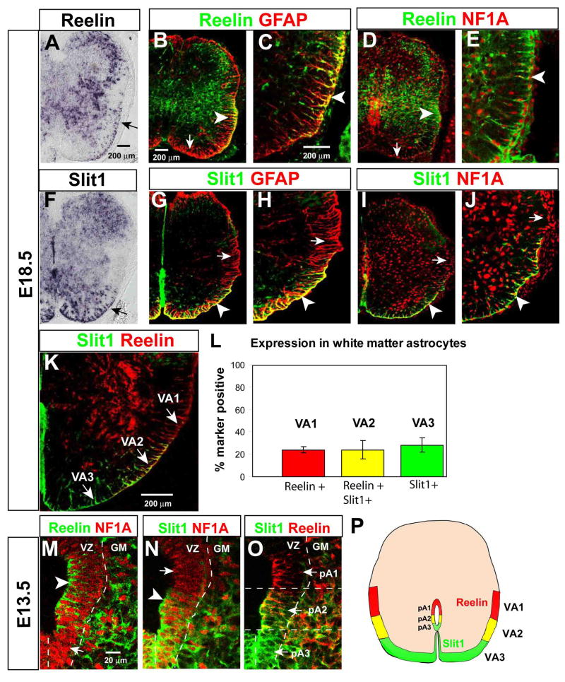
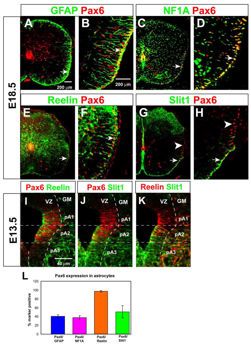
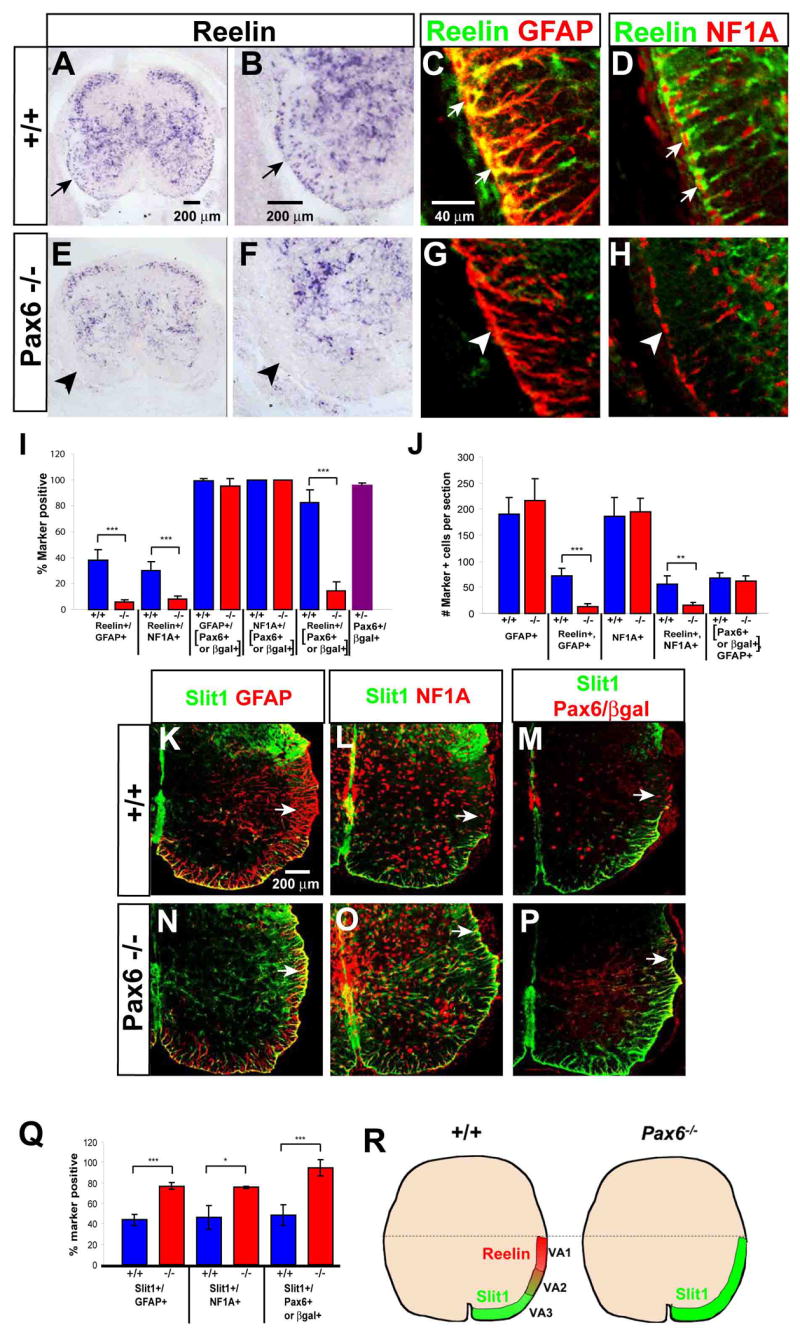
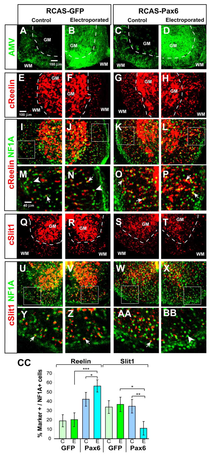
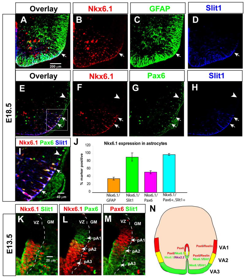
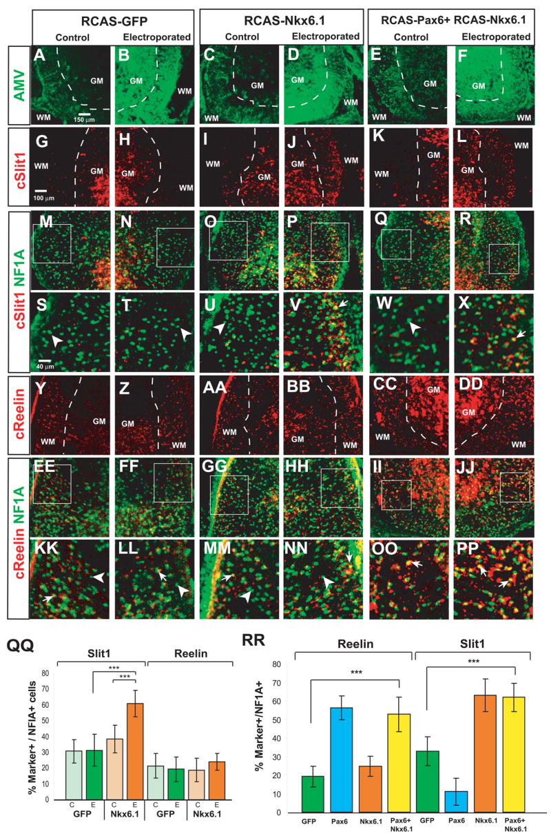
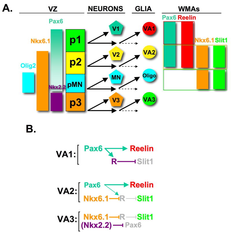
Astrocytes constitute the most abundant cell type in the central nervous system (CNS) and play diverse functional roles, but the ontogenetic origins of this phenotypic diversity are poorly understood. We have investigated whether positional identity, a fundamental organizing principle governing the generation of neuronal subtype diversity, is also relevant to astrocyte diversification. We identified three positionally distinct subtypes of white-matter astrocytes (WMA) in the spinal cord, which can be distinguished by the combinatorial expression of Reelin and Slit1. These astrocyte subtypes derive from progenitor domains expressing the homeodomain transcription factors Pax6 and Nkx6.1, respectively. Loss- and gain-of-function experiments indicate that the positional identity of these astrocyte subtypes is controlled by Pax6 and Nkx6.1 in a combinatorial manner. Thus, positional identity is an organizing principle underlying astrocyte, as well as neuronal, subtype diversification and is controlled by a homeodomain transcriptional code whose elements are reutilized following the specification of neuronal identity earlier in development.


Figures

Figure 1
Figure 1. Reelin and Slit1 define astrocyte subpopulations in the ventral spinal cord
(A, F) In situ hybridization for mouse Reelin and Slit1 mRNAs. Arrows indicate expression in ventral white matter. (B–E) Double-label immunohistochemistry for Reelin and GFAP (B–C) or NFIA (D–E). Arrowheads indicate double-positive cells in lateral white matter, arrows Reelin astrocytes in ventral white matter. (G–J) Double-label immunohistochemistry for Slit1-GFP and either GFAP (G–H) or NFIA (I–J), in Slit1GFP/+ embryos. Arrowheads indicate Slit1+ astrocytes in ventral white matter, arrows Slit1 astrocytes in the lateral white matter. (K) Double labeling for Reelin and Slit1-GFP reveals three subpopulations of ventral astrocytes (arrows). (L) Quantification of the percentage of NFIA+ astrocytes expressing each of the three markers. Data represent the mean±S.E.M. of 5–6 sections/embryo from 3 embryos. (M–O) Triple labeling for Reelin, Slit1-GFP and NFIA in the E13.5 ventral VZ, displayed as pairwise comparisons from the same section. Arrowheads and arrows in (M, N) indicate NFIA+ cells that are positive or negative, respectively, for Reelin (M) or Slit1 (N). (O) Overlay of Slit1 and Reelin reveals three adjacent progenitor domains in the VZ. (P) Composite schematic illustrating positions of VA1–VA3 astrocytes in the white matter at E18.5, and corresponding progenitor domains (pA1–3) in the VZ at E13.5.
Figure 2
Figure 2. Expression of Pax6 in Reelin+ astrocytes and their precursors
(A–H) Double-labeling for Pax6 (red) and the indicated markers (green) at E18.5. (B, D, F, and H) are higher magnification views of the areas indicated by arrows in (A, C, E and G), respectively. (I–K) Triple labeling for Pax6, Reelin, and Slit1-GFP in the E13.5 ventral VZ, displayed as pairwise comparisons from the same section. (L) Quantification of Pax6 expression among total (GFAP+ or NFIA+) astrocytes, and Reelin+ or Slit1+ WMAs.
Figure 3
Figure 3. Astrocyte subtype conversion in Pax6−/− mice
(A–B, E–F) In situ hybridization for Reelin mRNA in E18.5 spinal cord of wild-type (A–B) and Pax6 mutant (E–F) embryos. (B) and (F) are higher-magnification views of (A) and (E), respectively. Arrows in (A, B) indicate Reelin+ cells in the white matter. (C–D, G–H) Double-labeling for Reelin and GFAP (C, G) or NFIA (D, H) in wild-type (C–D) and Pax6 mutant (G–H) embryos. (I–J) Quantification of Reelin expression by WMAs in E18.5 wild type (blue bars) and Pax6−/− (red bars) spinal cord. . (I) The percentage of total GFAP+ or NFIA+ WMAs expressing Reelin is significantly reduced in the Pax6lacZ/lacZ mutant (***, p<.001). (J) The absolute number of GFAP+ and NFIA+ astrocytes in the white matter is not changed in the Pax6lacZ/lacZ spinal cord, nor is the average number of Pax6+ GFAP+ astrocytes. (*** and **, p<.001 and .002, respectively). . The data are derived from 5 wild type and 6 mutant embryos from 3 independent litters. (K–P) Spinal cord sections from Pax6+/+; Slit1GFP/+ (K–M) and Pax6lacZ/lacZ; Slit1GFP/+ (N–P) embryos double-labeled for the indicated markers. Arrows indicate VA1 astrocytes (K–M) that exhibit de-repression of Slit1 in the mutant (N–P). Red staining in (M, P) represents anti-Pax6 (M) or anti-βgal (P). (Q) Slit1 expression among total astrocytes (GFAP+ or NFIA+) is significantly increased in Pax6−/− embryos (*** and *, p = .0003, and .012, respectively). (H) Schematic illustrating the changes in Reelin and Slit1 expression by ventral astrocytes in the Pax6 mutant.
Figure 4
Figure 4. Pax6 is sufficient to promote Reelin expression and repress Slit1 expression in astrocytes
(A–D) Immunostaining for RCAS retroviral coat protein AMV in E12 spinal cord of chick embryos electroporated with either RCAS-GFP (A, B) or RCAS-Pax6 (C, D “WM,” white matter; “GM,” gray matter. (E-BB) Antibody staining for NFIA combined with fluorescent in situ hybridization for Reelin (E–P) or Slit1 (Q-BB) mRNAs. (I–L) and (U–X) are the same panels as in (E–H) and (Q–T), respectively, but include NF1A expression. (M–P) and (Y-BB) are higher power images of the boxed areas indicated in (I–L) and (U–X), respectively. Arrows indicate double-positive cells, arrowheads NFIA single-positive cells. (CC) The percentage of Reelin+ NFIA+ cells is significantly different between the electroporated (E) sides of Pax6 and GFP embryos (***, p <.001), as well as between the E and contralateral (C) sides of Pax6 embryos (*, p=.02). The reduction in Slit1 expression on the E side of Pax6 embryos is significant with respect to both the C side (**, p = .002), and the E side of GFP controls (*, p = .009). mean±S.E.M., n= 5 embryos per condition, two independent experiments.
Figure 5
Figure 5. Nkx6.1 is co-expressed by Slit1+ astrocytes and their precursors
(A–I) Triple-labeling for Nkx6.1, GFAP and Slit1-GFP (A–D) or Nkx6.1, Pax6 and Slit1-GFP (E–I). (I) is a higher power image of the boxed area in (E). Note that all Slit1+ cells in this domain (boxed area, E) are Nkx6.1+ and Pax6+ (E–G, I, arrows, white nuclei), while all Pax6+ cells dorsal to the boundary of Slit1 expression are Nkx6.1 (E–G, I, arrowheads). (J) Quantification of Nkx6.1 expression in astrocyte sub-populations. Nkx6.1 is expressed in > 90% of Slit1+ astrocytes (green bar). (K–M) Triple antibody labeling for Nkx6.1, Slit1-GFP and Pax6 in the E13.5 VZ, displayed as pairwise comparisons from the same section. The three progenitor domains are indicated (L, M, arrows). “GM,” gray matter. (N) Composite schematic illustrating relationship between the domains of Reelin and Slit1 expression, and those of Pax6 and Nkx6.1 expression, in the white matter at E18.5 and in the VZ at E13.5.
Figure 6
Figure 6. Nkx6.1 promotes Slit1 expression in astrocytes and overrides its repression by Pax6
Sections through the spinal cord of E12 chick embryos electroporated with the constructs indicated above the diagram, and labeled with the antibodies (AMV, NF1A) or cRNA probes (cSlit1, cReelin) indicated to the left. Abbreviations as in Fig 4. (M–R) and (EE-JJ) are the same panels as in (G–L) and (Y-DD), respectively, but include NFIA expression. (S–X) and (KK-PP) are higher power images of the boxed areas indicated in (M–R) and (EE-JJ), respectively. Arrows in (S–X) and (KK-PP) indicate double positive cells, arrowheads NFIA single positive cells. (QQ) The percentage of Slit1+ NFIA+ cells is significantly increased on the electroporated (E) side of Nkx6.1 embryos, relative to either the control (C ) side of the same embryos (***, p=.0003), or to the E side of GFP embryos (***, p<.001). (n=4 embryos per condition, two independent experiments). (RR) Bars represent mean ± S.E.M. from the E side only. The data from embryos electroporated with Pax6 alone (blue bars) or Nkx6.1 alone (orange bars) are reproduced from Figs. 4CC and 6QQ, respectively, and included for comparative purposes. The percentage of Slit1+NFIA+ cells in embryos mis-expressing both Nkx6.1 and Pax6 is significantly higher than in GFP controls (Slit1, ***, p<.001), and is similar to that in embryos expressing Nkx6.1 alone (QQ). (n=4 embryos per condition, 2 independent experiments).
Figure 7
Figure 7. A transcriptional code for astrocyte positional identity
(A) Schematic illustrating sequential generation of neurons and glia from different progenitor domains in the ventral spinal cord. VZ, ventricular zone; WMAs white matter astrocytes. The molecular phenotype of each of the 3 WMA subtypes is enclosed in a colored box. The three classes of WMAs are present in roughly equal proportions. Note that the expression boundaries of some of the transcription factors in the VZ shift from the neurogenic to the gliogenic phases; these shifts are omitted for simplicity. Note also that Nkx2.2, which is co-expressed with Nkx6.1 in the ventral-most (pA3) domain of the VZ during the gliogenic phase (Supplemental Figure S6), is not co-expressed with Nkx6.1 in VA3 WMAs (Supplemental Figure S7). (B) Model for the regulation of astrocyte identity by Pax6 and Nkx6.1. ”R” represents a hypothetical repressor of Slit1, which is activated by Pax6 and repressed by Nkx6.1. See Discussion for details. Repression of Pax6 by Nkx2.2 is likely important for the initial specification of VA3 identity within the VZ (see (A)), but does not contribute to the maintenance of VA3 identity in the WM.

References

    1. Bailey MS, Shipley MT. Astrocyte subtypes in the rat olfactory bulb: Morphological heterogeneity and differential laminar distribution. The Journal of comparative neurology. 1993;328:501–526. - PubMed
    1. Briscoe J, Ericson J. Specification of neuronal fates in the ventral neural tube. Current opinion in neurobiology. 2001;11:43–49. - PubMed
    1. Briscoe J, Pierani A, Jessell TM, Ericson J. A Homeodomain Protein Code Specifies Progenitor Cell Identity and Neuronal Fate in the Ventral Neural Tube. Cell. 2000;101:435–445. - PubMed
    1. Briscoe J, Sussel L, Serup P, Hartigan-O’Connor D, Jessell TM, Rubenstein JL, Ericson J. Homeobox gene Nkx2.2 and specification of neuronal identity by graded Sonic hedgehog signalling. Nature. 1999;398:622–627. - PubMed
    1. Brose K, Bland KS, Wang KH, Arnott D, Henzel W, Goodman CS, Tessier-Lavigne M, Kidd T. Slit proteins bind Robo receptors and have an evolutionarily conserved role in repulsive axon guidance. Cell. 1999;96:795–806. - PubMed

Publication types

MeSH terms