Comparative Study
. 2008 Jul 5;376(2):416-28.
doi: 10.1016/j.virol.2008.03.034. Epub 2008 May 5.

Virus growth and antibody responses following respiratory tract infection of ferrets and mice with WT and P/V mutants of the paramyxovirus Simian Virus 5

Affiliations
Comparative Study

Virus growth and antibody responses following respiratory tract infection of ferrets and mice with WT and P/V mutants of the paramyxovirus Simian Virus 5

Gerald A Capraro et al. Virology. .

Abstract

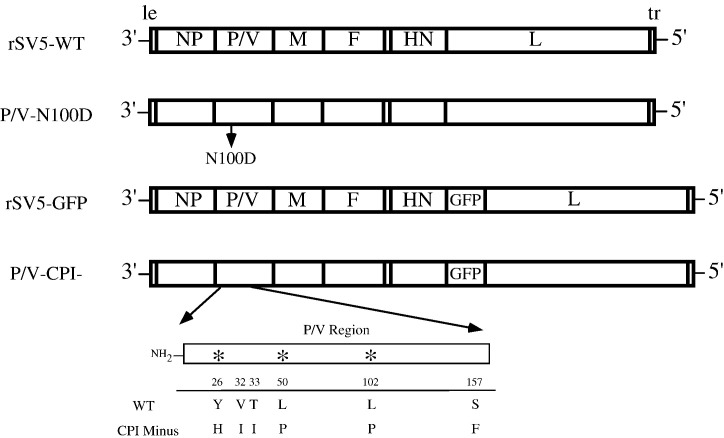
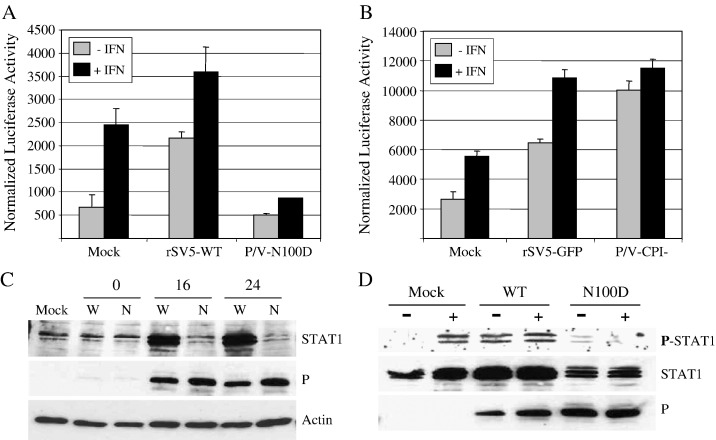
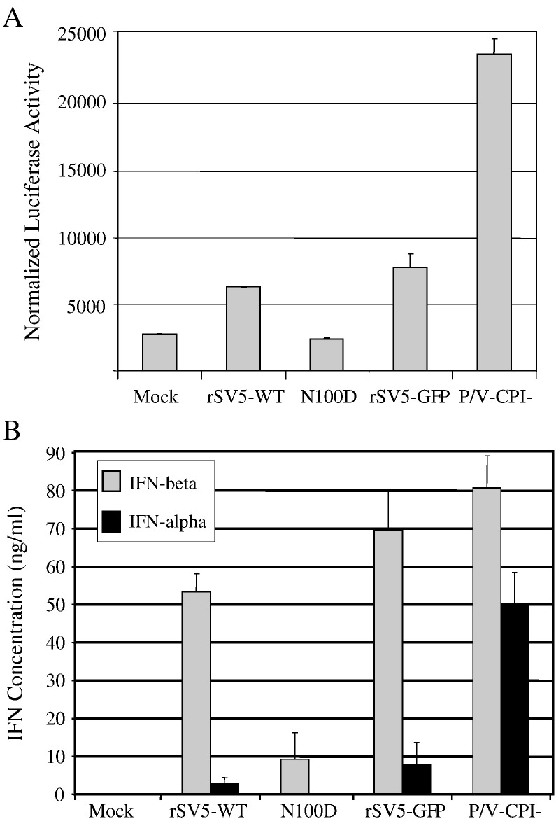
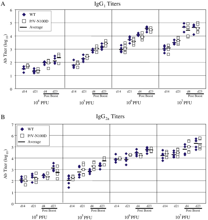
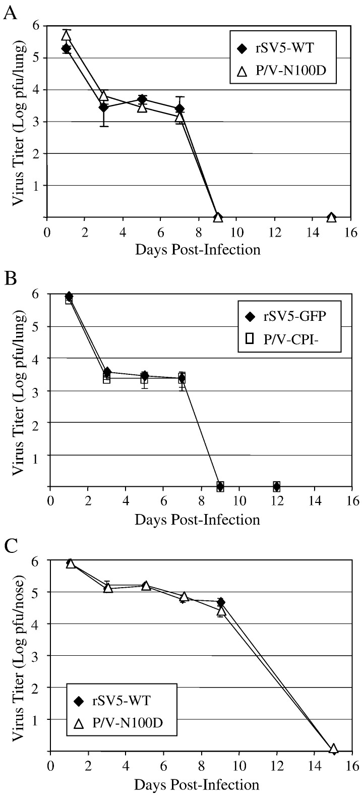
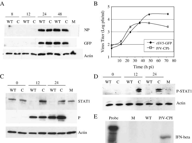
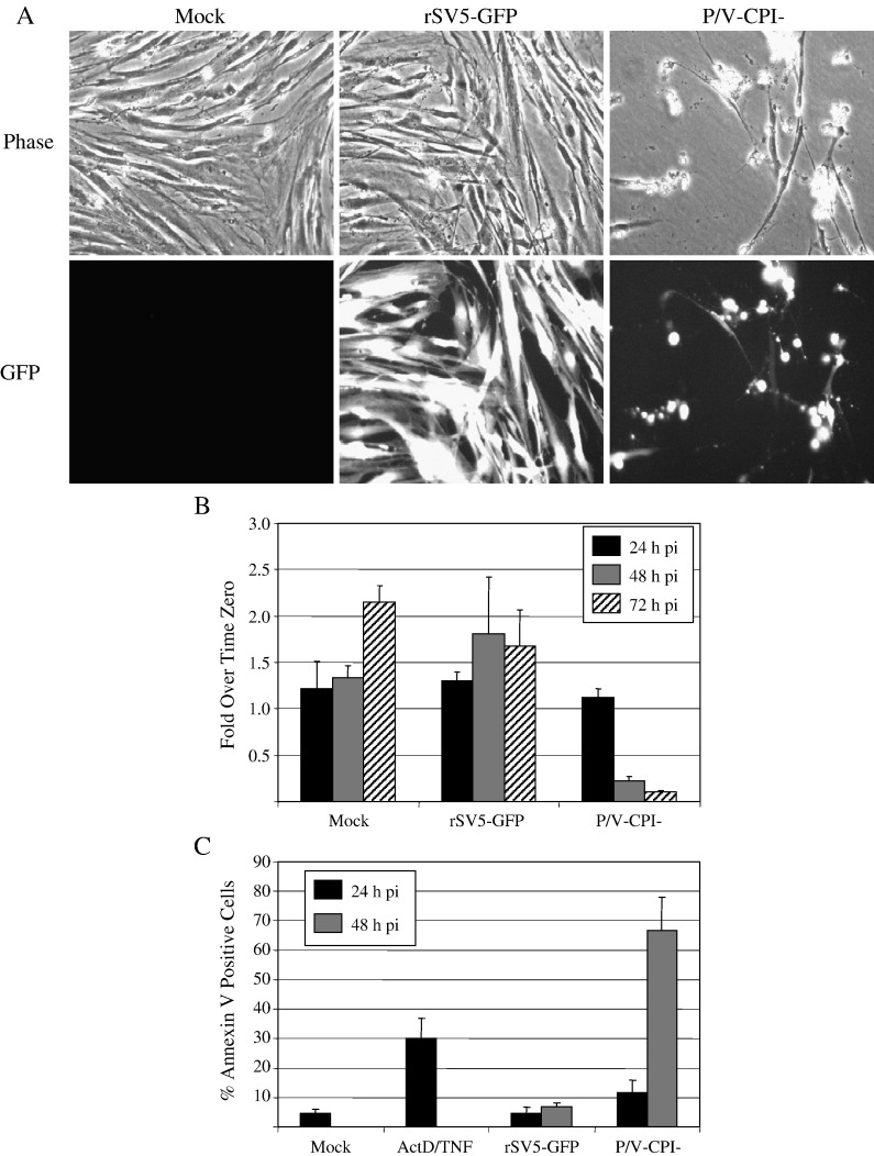
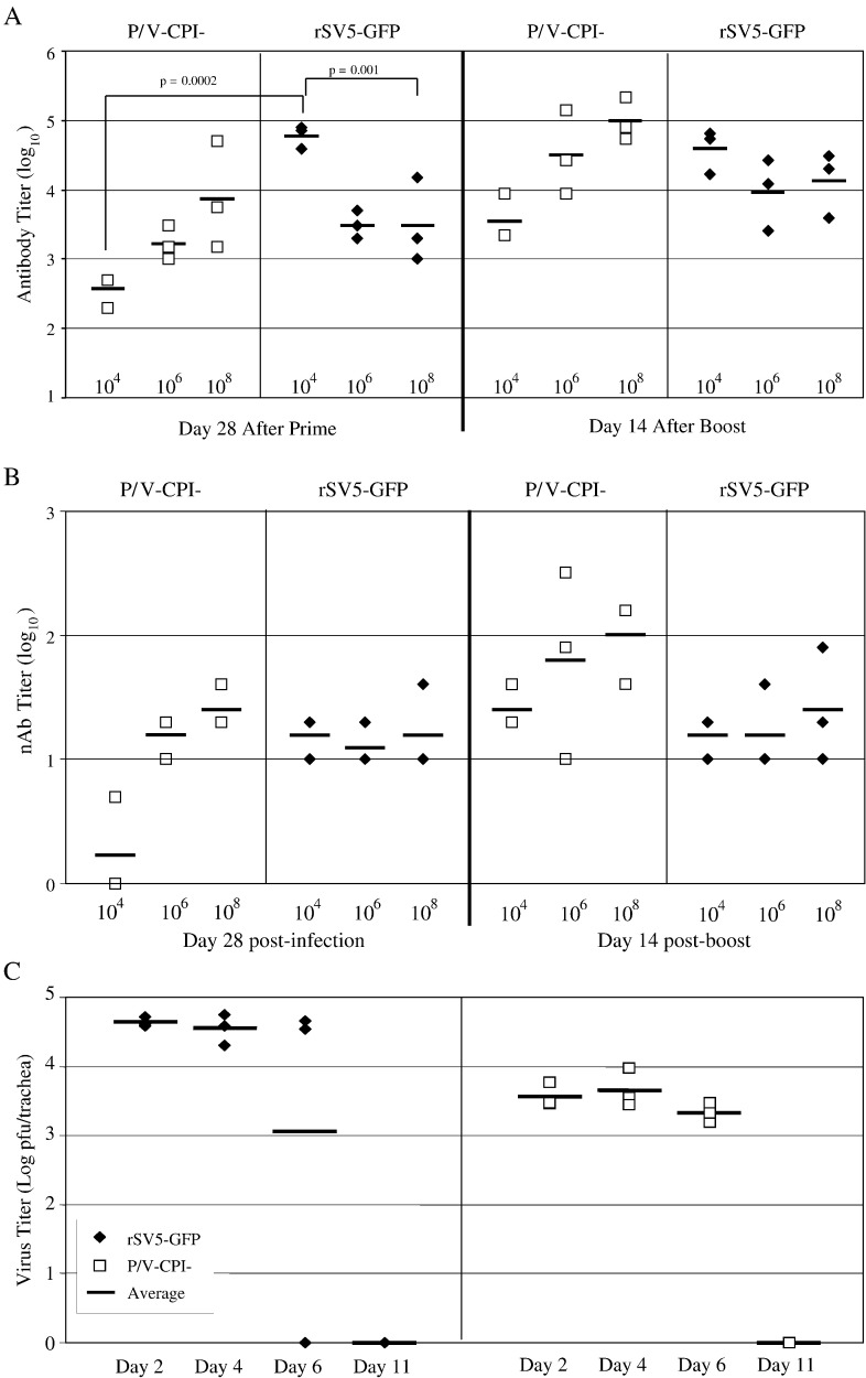
P/V gene substitutions convert the non-cytopathic paramyxovirus Simian Virus 5 (SV5), which is a poor inducer of host cell responses in human tissue culture cells, into a mutant (P/V-CPI-) that induces high levels of apoptosis, interferon (IFN)-beta, and proinflammatory cytokines. However, the effect of SV5-P/V gene mutations on virus growth and adaptive immune responses in animals has not been determined. Here, we used two distinct animal model systems to test the hypothesis that SV5-P/V mutants which are more potent activators of innate responses in tissue culture will also elicit higher antiviral antibody responses. In mouse cells, in vitro studies identified a panel of SV5-P/V mutants that ranged in their ability to limit IFN responses. Intranasal infection of mice with these WT and P/V mutant viruses elicited equivalent anti-SV5 IgG responses at all doses tested, and viral titers recovered from the respiratory tract were indistinguishable. In primary cultures of ferret lung fibroblasts, WT rSV5 and P/V-CPI- viruses had phenotypes similar to those established in human cell lines, including differential induction of IFN secretion, IFN signaling and apoptosis. Intranasal infection of ferrets with a low dose of WT rSV5 elicited approximately 500 fold higher anti-SV5 serum IgG responses compared to the P/V-CPI- mutant, and this correlated with overall higher viral titers for the WT virus in tracheal tissues. There was a dose-dependent increase in antibody response to infection of ferrets with P/V-CPI-, but not with WT rSV5. Together our data indicate that WT rSV5 and P/V mutants can elicit distinct innate and adaptive immunity phenotypes in the ferret animal model system, but not in the mouse system. We present a model for the effect of P/V gene substitutions on SV5 growth and immune responses in vivo.


Figures

Fig. 1
Fig. 1
Schematic diagram of SV5 viruses used in this study. The genome structures of rSV5-WT, P/V-N100D, WT rSV5-GFP and P/V-CPI− are shown schematically as negative sense RNA. Only rSV5-GFP and P/V-CPI− encode an additional gene (GFP) inserted between HN and L. The P/V-N100D and P/V-CPI− mutants encode a single N100D mutation or a set of six CPI− mutations in the shared P/V N-terminal region, respectively. The differences between the WT and CPI− P/V region is shown at the bottom with stars denoting the residues shown previously to be important for targeting STAT1 for degradation (Chatziandreou et al., 2002). le, leader; tr, trailer.
Fig. 2
Fig. 2
WT and P/V mutant SV5 with differential ability to block IFN signaling in mouse cells. (A and B) Differential activation of ISRE-luciferase. Mouse CL7 cells were co-transfected with pSV-betagal and a plasmid containing the luciferase gene under control of an ISRE. Twenty-four hours post-transfection, cells were mock infected or infected at an moi of 20 with WT rSV5 or rSV5-P/V-N100D (panel A) or with rSV5-GFP or rSV5-P/V-CPI− (panel B). At 15 h pi, cells were incubated for 6 h with (black bars) or without (grey bars) 1000 U of mouse IFN-beta, after which lysates were prepared and luciferase and Beta-gal assays were performed. Normalized luciferase activities are representative of three independent experiments with bars representing standard deviation from the mean. C) STAT1 levels in P/V-N100D infected cells. CL7 cells were mock infected or infected at high moi with rSV5-WT (W lanes) or P/V-N100D (N lanes). Cell lysates were prepared at the indicated times pi and analyzed by Western blotting for levels of STAT1, viral P protein and cellular actin. D) STAT1 phosphorylation. CL7 cells were mock infected or infected at high moi with the indicated viruses. At 24 h pi, cells were treated with (+) or without (−) 1000 U of mouse IFN-beta for 6 h. Cell lysates were analyzed for STAT1, P protein and STAT1 phosphorylated on tyrosine 701 (P-STAT1).
Fig. 3
Fig. 3
Differential induction of IFN in mouse cells infected with WT rSV5 and P/V mutants. A) Induction of the IFN-β promoter. CL7 cells were co-transfected with pSV-betagal and a plasmid containing the luciferase gene under control of the IFN-beta promoter. Twenty-four hours post-transfection, cells were mock infected or infected at high moi with the indicated viruses. Cells were lysed at 15 h pi, and normalized luciferase activity was calculated as in Fig. 2. Data are the average of three experiments with bars indicating standard deviation from the mean. B) IFN ELISA. CL7 cells were infected at high moi with the indicated viruses, and media collected at 24 h pi were analyzed by ELISA for levels of IFN-beta (grey bars) or IFN-alpha (black bars). Results are the average of three experiments with bars indicating standard deviation from the mean.
Fig. 4
Fig. 4
Multicycle growth of WT and P/V mutant SV5 in mouse CL7 cells. Mouse CL7 cells were infected at an moi of 0.05 with the indicated WT rSV5 or P/V mutants. At the indicated times pi, media were collected and assayed for infectious virus by plaque assay. Data are the average of three independent experiments.
Fig. 5
Fig. 5
Effect of virus dose on antibody responses in mice infected with WT SV5 and P/V-N100D. Groups of 5 BALB/c mice were infected i.n. with the indicated doses (in pfu) of WT rSV5 (closed diamonds) or P/V-N100D (open squares). At days 14 and 21 pi sera were collected and assayed by ELISA for anti-SV5 IgG1 (panel A) and IgG2a (panel B) responses. Mice were boosted with an equivalent amount of homologous virus, and levels of anti-SV5 antibodies were determined at day 4 and day 23 post boost. Each symbol represents data for an individual mouse with the horizontal bar indicating the mean value for the group.
Fig. 6
Fig. 6
Growth of WT and P/V mutant rSV5 in mice. Groups of BALB/c mice were infected i.n. with 106 pfu of rSV5-WT or P/V-N100D (panels A and C) or with rSV5-GFP or P/V-CPI− (panel B). At the indicated days pi, lungs (panels A and B) or nasal tissue (panel C) were harvested and assayed for infectious virus by plaque assay as described in Materials and methods. Data are the average of three mice per time point, with bars representing the standard deviation from the mean.
Fig. 7
Fig. 7
In vitro growth properties of rSV5-GFP and P/V-CPI− mutant in ferret lung fibroblast cells. A) GFP expression. Primary cultures of ferret fibroblast cells were mock infected or infected at an moi of 10 with rSV5-GFP or P/V-CPI−. Cell lysates prepared at the indicated times pi were analyzed by western blotting for viral NP and GFP or for cellular actin. B) Low moi growth analysis. Ferret cells were infected with rSV5-GFP or P/V-CPI− at an moi of 0.05. Media were collected at the indicated times pi for analysis by plaque assay. Data are representative of four independent experiments. C and D) STAT1 status in ferret fibroblasts. Primary cultures of ferret fibroblasts were mock infected (M lane) or infected at an moi of 10 with WT rSV5-GFP (WT lanes) or the P/V-CPI− mutant (C lanes). At the indicated times pi, cell lysates were prepared for analysis by Western blotting for cellular STAT1, STAT1 phosphorylated on tyrosine 701 (P-STAT1), viral P protein or cellular actin as indicated. E) IFN-β gene expression. RNA was harvested at 20 h pi from ferret fibroblasts that were mock infected (M lane) or infected at an moi of 10 with rSV5-GFP (WT lanes) or P/V-CPI− and analyzed by RPA using a 32P-radiolabeled anti-sense riboprobe (P lane) specific for ferret IFN-beta. Results are representative of three independent experiments.
Fig. 8
Fig. 8
Induction of CPE and apoptotic markers following infection of ferret fibroblasts with the P/V-CPI− mutant but not rSV5-GFP. A) Ferret fibroblasts were mock infected or infected at an moi of 10 with WT rSV5-GFP or the P/V-CPI− mutant. Cells were examined by microscopy at 48 hours pi for CPE and GFP expression. B) The number of viable cells at the indicated times pi was determined by trypan blue exclusion. Data are expressed as fold change in viable cell number compared to the starting number of cells and are the average of 3 samples per timepoint. Bars represent standard deviation from the mean. C) Cells were analyzed by flow cytometry at the indicated times pi for staining with PE-conjugated annexin V. Data are the average of 3 samples per timepoint, with bars representing the standard deviation from the mean.
Fig. 9
Fig. 9
Antibody response and viral titers elicited by intranasal infection of ferrets with rSV5-GFP and P/V-CPI−. A and B) ELISA and neutralizing antibody titers. Groups of 3 ferrets each were infected i.n. with 104, 106, or 108 pfu of rSV5-GFP or P/V-CPI−. At day 28 pi, serum was collected and ferrets were boosted with an equivalent amount of homologous virus. Levels of anti-SV5 antibodies in serum from day 28 and d14 post boost (panel A) and neutralizing titers in d28 and d14 post boost serum (panel B) were determined as described in Materials and methods. Each symbol represents data for an individual ferret with the horizontal bars indicating the mean value for the group. Results are representative of two independent experiments. C) Viral titers. Ferrets were infected i.n. with 104 pfu of rSV5-GFP or P/V-CPI−, and viral titers in the upper trachea of animals were determined at the indicated days pi. Each symbol represents the titer for one experimental animal, and the bar represents the mean value for the group.

Similar articles

Cited by

References

    1. Andrejeva J., Poole E., Young D.F., Goodbourn S., Randall R.E. The p127 subunit (DDB1) of the UV-DNA damage repair binding protein is essential for the targeted degradation of STAT1by the V protein of the paramyxovirus Simian Virus 5. J. Virol. 2002;76:11379–11386. - PMC - PubMed
    1. Andrejeva J., Childs K.S., Young D.F., Carlos T.S., Stock N., Goodbourn S., Randall R.E. The V proteins of paramyxoviruses bind the IFN-inducible RNA helicase, mda-5, and inhibit its activation of the IFN-beta promoter. Proc. Natl. Acad. Sci. U. S. A. 2004;101(49):17264–17269. - PMC - PubMed
    1. Arimilli S., Alexander-Miller M.A., Parks G.D. A Simian Virus 5 (SV5) P/V mutant is less cytopathic than wild-type SV5 in human dendritic cells and is a more effective activator of dendritic cell maturation and function. J. Virol. 2006;80:3416–3427. - PMC - PubMed
    1. Arimilli S., Johnson J.B., Alexander-Miller M.A., Parks G.D. TLR-4 and -6 agonists reverse apoptosis and promote maturation of Simian Virus 5 infected human dendritic cells through NFkB-dependent pathways. Virology. 2007;365:144–156. - PMC - PubMed
    1. Atmar R.L., Keitela W.A., Catea T.R., Munozb F.M., Rubend F., Coucha R.B. A dose-response evaluation of inactivated influenza vaccine given intranasally and intramuscularly to healthy young adults. Vaccine. 2007;25(29):5367–5373. - PMC - PubMed

Publication types

MeSH terms