Comparative Study
. 2008 Jul;295(1):H10-20.
doi: 10.1152/ajpheart.01277.2007. Epub 2008 May 2.

Sex differences in circulating and renal angiotensins of hypertensive mRen(2). Lewis but not normotensive Lewis rats

Affiliations
Comparative Study

Sex differences in circulating and renal angiotensins of hypertensive mRen(2). Lewis but not normotensive Lewis rats

Karl D Pendergrass et al. Am J Physiol Heart Circ Physiol. 2008 Jul.

Abstract

Sex differences in blood pressure are evident in experimental models and human subjects, yet the mechanisms underlying this disparity remain equivocal. The current study sought to define the extent of male-female differences in the circulating and tissue renin-angiotensin aldosterone systems (RAASs) of congenic mRen(2). Lewis and control Lewis rats. Male congenics exhibited higher systolic blood pressure than females [200 +/- 4 vs. 146 +/- 7 mmHg, P < 0.01] or Lewis males and females [113 +/- 2 vs. 112 +/- 2 mmHg, P > 0.05]. Plasma ANG II levels were twofold higher in male congenics [47 +/- 3 vs. 19 +/- 3 pM, P < 0.01] and fivefold higher than in male or female Lewis rats [6 +/- 1 vs. 6 +/- 1 pM]. ANG I levels were also highest in the males; however, plasma ANG-(1-7) was higher in female congenics. Male congenics exhibited greater circulating renin and angiotensin-converting enzyme (ACE) activities, as well as angiotensinogen, than female littermates. Renal cortical and medullary ANG II levels were also higher in the male congenics versus all the other groups; ANG I was lower in the males. Cortical ACE2 activity was higher in male congenics, yet neprilysin activity and protein were greater in the females, which may contribute to reduced renal levels of ANG II. These data reveal that sex differences in both the circulating and renal RAAS are apparent primarily in the hypertensive group. The enhanced activity of the RAAS in male congenics may contribute to the higher pressure and tissue injury evident in the strain.


Figures
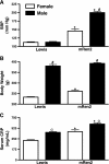
Fig. 1.
Fig. 1.
Cardiovascular indexes in Lewis and mRen(2).Lewis rats. A: systolic blood pressure (SBP, in mmHg) was measured by tail-cuff in conscious Lewis and hypertensive mRen(2).Lewis (mRen2) rats. B: body weight (in g). C: complement-reactive peptide (CRP) was determined in the serum from male and female Lewis and mRen(2).Lewis rats. Values are means ± SE. #P < 0.001 between sex differences; αP < 0.01 between sex differences; *P < 0.001 between strains (n = 5 to 6 rats/group).
Fig. 2.
Fig. 2.
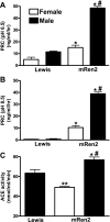
Circulating renin-angiotensin aldosterone system hormones in Lewis and mRen(2).Lewis rats. Plasma angiotensins and aldosterone were measured by separate radioimmunoassays for ANG II (A), ANG I (B), ANG-(1-7) (C), and aldosterone (D). Peptide values are expressed as picomolar concentration. Plasma aldosterone is expressed as nanograms per deciliter. Values are means ± SE. #P < 0.001 between sex differences; *P < 0.001 between strains; ɛP < 0.05 between strains (n = 5 to 6 rats/group).
Fig. 3.
Fig. 3.
Representative immunoblot of plasma angiotensinogen in the Lewis and mRen(2).Lewis rats using a COOH-terminal antibody. Angiotensinogen was determined in plasma (0.38 μl) from the male Lewis (ML), female Lewis (FL), male mRen(2).Lewis (MC), and the female mRen(2).Lewis (FC) rats. Quantification of the bands: protein expression was quantified by densitometry [in absorbance units (AU)] for the 55/60-kDa bands, respectively. OD, optical density. Values are means ± SE. *P < 0.01 between strains (n = 4 rats/group).
Fig. 4.
Fig. 4.
Circulating renin and angiotensin-converting enzyme (ACE) activities in the Lewis and mRen(2).Lewis rats. A: plasma renin concentration (PRC) was measured at the pH optima for rat renin (pH of 6.5). B: PRC was measured at the pH optima of mouse remain (pH of 8.5). C: serum ACE activity was measured using the synthetic substrate 3H-[Hip-His-Leu]. PRCs and ACE activity were expressed as nanograms per milliliters per hour and nanomoles per milliliters per minute, respectively. Values are means ± SE. #P < 0.001 between sex differences; *P < 0.001 between strains; **P < 0.001 compared with male Lewis rats (n = 5 rats/group).
Fig. 5.
Fig. 5.
Renal indexes in the Lewis and mRen(2).Lewis rats. A: renal hypertrophy was expressed as the ratio of left kidney to body weight (in mg/g). B: proteinuria was measured from a 24-h collection of urine (in mg protein·kg−1·day−1). C: urinary creatinine excretion (in mg·kg−1·day−1). D: urinary 8-hydroxy-2′-deoxyguanosine (8-OH-dG) excretion (in μg·kg−1·day−1). Values are means ± SE. #P < 0.001 between sex differences; *P < 0.001 between strains (n = 5 rats/group).
Fig. 6.
Fig. 6.
Renal cortical angiotensins in the Lewis and mRen(2).Lewis rats. Angiotensins were measured by separate radioimmunoassays of cortical extracts and expressed (in fmol/mg protein) for ANG II (A), ANG I (B), and ANG-(1-7) (C). Values are means ± SE. #P < 0.001 between sex differences; ɛP < 0.05 between strains (n = 4–6 rats/group).
Fig. 7.
Fig. 7.
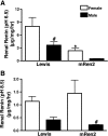
Renal renin concentrations in the Lewis and mRen(2).Lewis rats. A: renal renin concentration was measured at the pH optima for rat renin (pH of 6.5). B: renal renin concentration was measured at the pH optima of mouse renin (pH of 8.5). Renal renin concentrations were expressed (in μg·mg protein−1·h−1). Values are means ± SE. #P < 0.05 between sex differences; *P < 0.01 between strains (n = 3 to 4 rats/group).
Fig. 8.
Fig. 8.
Renal enzymatic activities in the Lewis and mRen(2).Lewis rats. A: ACE activity. B: ACE2 activity. C and D: neprilysin (ANG I and ANG II as substrates) activities were determined in solubilized cortical membranes. Renal enzyme activities were expressed (in nmol·mg protein−1·min−1) for ACE and (in fmol·mg protein−1·min−1) for ACE2 and neprilysin. Peptidase values are means ± SE. #P < 0.01 between sex differences; *P < 0.01 between strains (n = 4 rats/group).
Fig. 9.
Fig. 9.
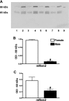
Immunoblot analysis of cortical neprilysin in male and female mRen(2).Lewis rats. Solubilized cortical membranes (10 μg) were separated by SDS-PAGE and reacted against a neprilysin antibody. A: for the full-length gel, lanes 1, 3, 5, and 7 are male cortical extracts, lanes 2, 4, 6, and 8 are female extracts, and lane 9 is the neprilysin standard (88 kDa). B and C: neprilysin protein expression was quantified by densitometry (in AU) for the 88- and 65-kDa bands, respectively. Values are means ± SE. #P < 0.01 (n = 4 rats/group).
Fig. 10.
Fig. 10.
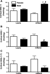
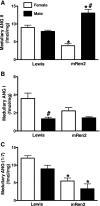
Renal medullary angiotensins in Lewis and mRen(2).Lewis rats. Angiotensins were measured by separate radioimmunoassays and expressed (in fmol/mg protein) for ANG II (A), ANG I (B), and ANG-(1-7) (C). Values are means ± SE. #P < 0.01 between sex differences; *P < 0.01 between strains (n = 5 rats/group).
Fig. 11.
Fig. 11.
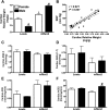
Cardiac indexes in the Lewis and mRen(2).Lewis rats. A: cardiac hypertrophy was expressed as the ratio of the left ventricle to body weight (in mg/g). B: association of cardiac hypertrophy and the blood pressure data. C: cardiac ANG II. D: cardiac ANG-(1-7). Angiotensin peptides were measured by separate radioimmunoassays (in fmol/mg protein; n = 4–6 rats/group). E and F: ACE and ACE2 activities were determined in solubilized cardiac membranes. Renal enzyme activities were expressed (in nmol·mg protein−1·min−1) for ACE and (in fmol·mg protein−1·min−1) for ACE2. Peptidase values are means ± SE. #P < 0.01 between sex differences; *P < 0.01 between strains (n = 4 rats/group).

References

    1. Allred AJ, Chappell MC, Ferrario CM, Diz DI. Differential actions of renal ischemic injury on the intrarenal angiotensin system. Am J Physiol Renal Physiol 279: F636–F645, 2000. - PubMed
    1. Allred AJ, Diz DI, Ferrario CM, Chappell MC. Pathways for angiotensin-(1-7) metabolism in pulmonary and renal tissues. Am J Physiol Renal Physiol 279: F841–F850, 2000. - PubMed
    1. Armando I, Jezova M, Juorio AV, Terron JA, Falcon-Neri A, Semino-Mora C, Imboden H, Saavedra JM. Estrogen upregulates renal angiotensin II AT2 receptors. Am J Physiol Renal Physiol 283: F934–F943, 2002. - PubMed
    1. August P, Oparil S. Hypertension in women. J Clin Endocrinol Metab 84: 1862–1866, 1999. - PubMed
    1. Bachmann J, Feldmer M, Ganten U, Stock G, Ganten D. Sexual dimorphism of blood pressure: possible role of the renin-angiotensin system. J Steroid Biochem 40: 511–515, 1991. - PubMed

Publication types

MeSH terms