. 2008 Jul;190(13):4666-76.
doi: 10.1128/JB.00093-08. Epub 2008 May 2.

Mutagenesis of the Shigella flexneri autotransporter IcsA reveals novel functional regions involved in IcsA biogenesis and recruitment of host neural Wiscott-Aldrich syndrome protein

Affiliations

Mutagenesis of the Shigella flexneri autotransporter IcsA reveals novel functional regions involved in IcsA biogenesis and recruitment of host neural Wiscott-Aldrich syndrome protein

Kerrie L May et al. J Bacteriol. 2008 Jul.

Abstract

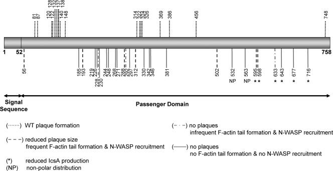
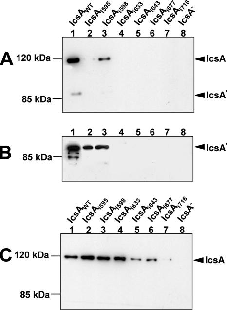
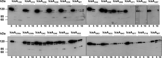
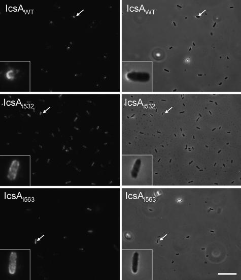
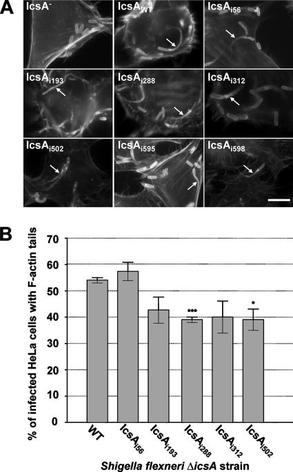
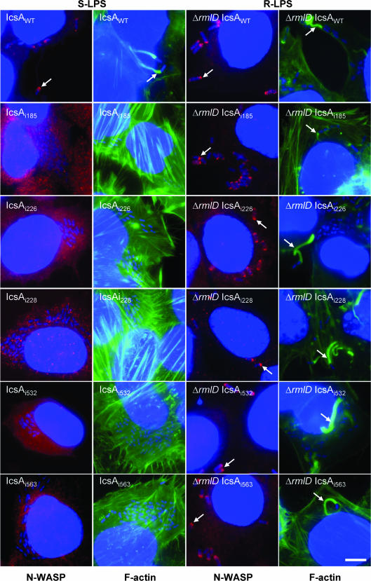
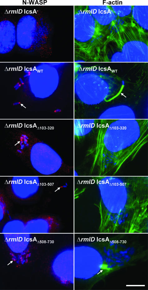
The IcsA (VirG) protein of Shigella flexneri is a polarly localized, outer membrane protein that is essential for virulence. Within host cells, IcsA activates the host actin regulatory protein, neural Wiskott-Aldrich syndrome protein (N-WASP), which in turn recruits the Arp2/3 complex, which nucleates host actin to form F-actin comet tails and initiate bacterial motility. Linker insertion mutagenesis was undertaken to randomly introduce 5-amino-acid in-frame insertions within IcsA. Forty-seven linker insertion mutants were isolated and expressed in S. flexneri Delta icsA strains. Mutants were characterized for IcsA protein production, cell surface expression and localization, intercellular spreading, F-actin comet tail formation, and N-WASP recruitment. Using this approach, we have identified a putative autochaperone region required for IcsA biogenesis, and our data suggest an additional region, not previously identified, is required for N-WASP recruitment.


Figures

FIG. 1.
FIG. 1.
Schematic locations of the 5 amino acid insertions in 47 mutant IcsAi proteins and the corresponding phenotypes. The location of the linker insertion for each of the 47 IcsAi mutants is indicated with a vertical line.
FIG. 2.
FIG. 2.
Western blot analyses of IcsAi mutant production and secretion. (A) Whole-cell lysates from log-phase S. flexneri ΔicsA strains expressing IcsAWT or IcsAi mutants. (B) Tricholoroacetic acid-precipitated culture supernatants from log-phase S. flexneri ΔicsA strains expressing IcsAWT or IcsAi mutants. (C) Whole-cell lysates from E. coli UT5600 strains expressing IcsAWT or IcsAi mutants. All samples were electrophoresed on a 7.5% SDS-PAGE gel prior to Western blot analysis with anti-IcsA antibodies. The 116-kDa band corresponds to full-length IcsA, and the 95-kDa band corresponds to the cleaved form (IcsA′). Samples represent 1 × 10⁸ cells or the culture supernatant equivalent of 1 × 10⁹ cells.
FIG. 3.
FIG. 3.
Trypsin accessibility of IcsAi mutants. Log-phase cultures of E. coli UT5600 expressing IcsAWT or IcsAi mutants were treated with 0.1 μg/ml of trypsin at 25°C. Aliquots were taken at 0 min, 5 min, and 10 min and supplemented with 1 mM phenylmethylsulphonylfluoride to inhibit further proteolysis. Whole-cell lysates were electrophoresed on a 7.5% SDS-PAGE gel and subjected to Western blot analysis with anti-IcsA antibody. Samples represent the equivalent of 1 × 10⁹ cells.
FIG. 4.
FIG. 4.
Nonpolar distribution of IcsAi532 and IcsAi563 mutants on the surface of S. flexneri. IF microscopy of IcsA surface distribution. Log-phase cultures of S. flexneri strains expressing either IcsAWT or IcsAi were formalin fixed and labeled with anti-IcsA antibodies and Alexa 488-conjugated goat antirabbit secondary antibodies. For clarity, the IF image for each strain is accompanied by an overlay of the IF image with the corresponding phase-contrast image. Bar = 10 μm.
FIG. 5.
FIG. 5.
F-actin comet tail formation by intracellular S. flexneri ΔicsA strains expressing IcsA-i mutants. (A) IF microscopy of F-actin tail formation by intracellular S. flexneri ΔicsA strain expressing IcsA-i mutants. HeLa cells infected with S. flexneri were labeled with anti-LPS antibodies and Alexa 594-conjugated donkey antirabbit antibodies, and F-actin was labeled with FITC-phalloidin. Arrows indicate F-actin tail formation. Strains were assessed in three independent experiments. Scale bar = 10 μm. (B) Frequency of F-actin tail formation by S. flexneri ΔicsA strains expressing IcsA-i mutants. HeLa cells were infected with S. flexneri strains and examined by IF microscopy as detailed in Materials and Methods. The frequency of F-actin tail formation was determined by observing the percentage of infected HeLa cells (n = 100) that had at least 1 F-actin tail. Data represent means ± standard errors. *, P < 0.05; ***, P < 0.001 (determined by Student's unpaired two-tailed t test). Data are from three independent experiments.
FIG. 6.
FIG. 6.
N-WASP recruitment and F-actin comet tail formation by intracellular S. flexneri strains expressing the IcsAi proteins with either smooth or rough LPS. IF microscopy of F-actin tail formation and N-WASP recruitment by intracellular S. flexneri strains expressing IcsA-i mutants. HeLa cells were infected with S. the flexneri ΔicsA (S-LPS) or S. flexneri ΔicsA ΔrmlD (R-LPS) strain expressing IcsAi proteins and formalin fixed. Bacteria were labeled with DAPI (blue), F-actin was labeled with FITC-phalloidin (green), and N-WASP was labeled with anti-N-WASP and Alex 594-conjugated donkey antirabbit antibodies (red) as detailed in Materials and Methods. Arrows indicate F-actin tail formation and N-WASP recruitment. Strains were assessed in two independent experiments. Bar = 10 μm.
FIG. 7.
FIG. 7.
N-WASP recruitment and F-actin comet tail formation by intracellular S. flexneri strains expressing IcsA deletion mutant proteins with either smooth or rough LPS. IF microscopy of F-actin tail formation and N-WASP recruitment by intracellular S. flexneri strains expressing IcsAΔ mutants. HeLa cells were infected with S. flexneri ΔicsA (S-LPS) or S. flexneri ΔicsA ΔrmlD (R-LPS) strains expressing IcsA deletion mutant proteins and formalin fixed. Bacteria were labeled with DAPI (blue), F-actin was labeled with FITC-phalloidin (green), and N-WASP was labeled with anti-N-WASP and Alex 594-conjugated donkey antirabbit antibodies (red) as detailed in Materials and Methods. Arrows indicate F-actin tail formation or N-WASP recruitment. Strains were assessed in two independent experiments. Bar = 10 μm.

Similar articles

Cited by

References

    1. Baker, S., J. Gunn, and R. Morona. 1999. The Salmonella typhi melittin resistance gene pqaB affects intracellular growth in PMA-differentiated U937 cells, polymyxin B resistance and lipopolysaccharide. Microbiology 145367-378. - PubMed
    1. Bernardini, M. L., J. Mounier, H. d'Hauteville, M. Coquis-Rondon, and P. J. Sansonetti. 1989. Identification of icsA, a plasmid locus of Shigella flexneri that governs bacterial intra- and intercellular spread through interaction with F-actin. Proc. Natl. Acad. Sci. USA 863867-3871. - PMC - PubMed
    1. Berthiaume, F., N. Rutherford, and M. Mourez. 2007. Mutations affecting the biogenesis of the AIDA-I autotransporter. Res. Microbiol. 158348-354. - PubMed
    1. Brahmbhatt, H. N., A. A. Lindberg, and K. N. Timmis. 1992. Shigella lipopolysaccharide: structure, genetics, and vaccine development. Curr. Top. Microbiol. Immunol. 18045-64. - PubMed
    1. Brandon, L. D., N. Goehring, A. Janakiraman, A. W. Yan, T. Wu, J. Beckwith, and M. B. Goldberg. 2003. IcsA, a polarly localized autotransporter with an atypical signal peptide, uses the Sec apparatus for secretion, although the Sec apparatus is circumferentially distributed. Mol. Microbiol. 5045-60. - PubMed

Publication types

MeSH terms

LinkOut - more resources