. 2008 Jul;190(13):4489-500.
doi: 10.1128/JB.00414-08. Epub 2008 May 2.

Temperature sensitivity and cell division defects in an Escherichia coli strain with mutations in yghB and yqjA, encoding related and conserved inner membrane proteins

Affiliations

Temperature sensitivity and cell division defects in an Escherichia coli strain with mutations in yghB and yqjA, encoding related and conserved inner membrane proteins

Kandi Thompkins et al. J Bacteriol. 2008 Jul.

Abstract

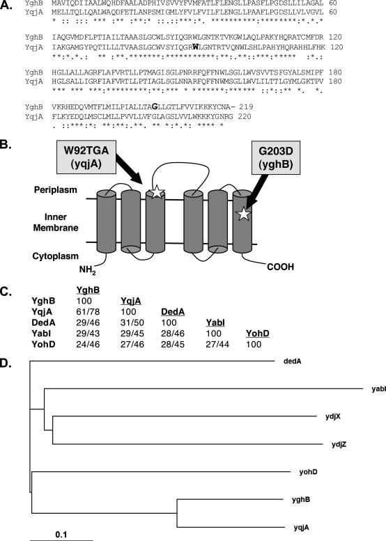
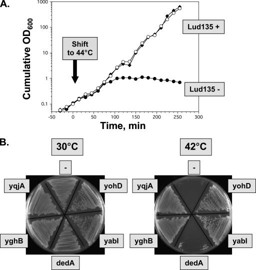
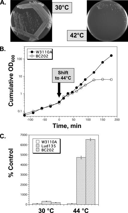
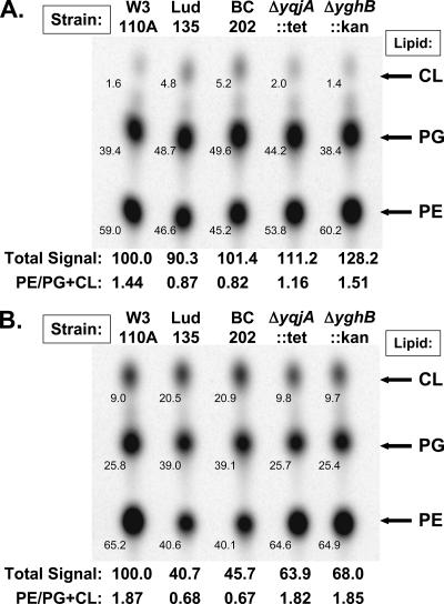
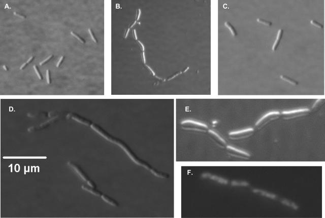
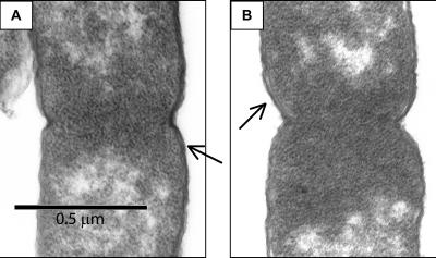
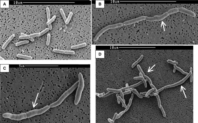
Ludox density gradients were used to enrich for Escherichia coli mutants with conditional growth defects and alterations in membrane composition. A temperature-sensitive mutant named Lud135 was isolated with mutations in two related, nonessential genes: yghB and yqjA. yghB harbors a single missense mutation (G203D) and yqjA contains a nonsense mutation (W92TGA) in Lud135. Both mutations are required for the temperature-sensitive phenotype: targeted deletion of both genes in a wild-type background results in a strain with a similar phenotype and expression of either gene from a plasmid restores growth at elevated temperatures. The mutant has altered membrane phospholipid levels, with elevated levels of acidic phospholipids, when grown under permissive conditions. Growth of Lud135 under nonpermissive conditions is restored by the presence of millimolar concentrations of divalent cations Ca(2+), Ba(2+), Sr(2+), or Mg(2+) or 300 to 500 mM NaCl but not 400 mM sucrose. Microscopic analysis of Lud135 demonstrates a dramatic defect at a late stage of cell division when cells are grown under permissive conditions. yghB and yqjA belong to the conserved and widely distributed dedA gene family, for which no function has been reported. The two open reading frames encode predicted polytopic inner membrane proteins with 61% amino acid identity. It is likely that YghB and YqjA play redundant but critical roles in membrane biology that are essential for completion of cell division in E. coli.


Figures

FIG. 1.
FIG. 1.
The E. coli DedA family of related predicted IM proteins. (A) Amino acid alignment between YqjA and YghB. W92 and G203, the sites of mutations in Lud135 in YqjA and YghB, respectively, are shown in bold. (B) Predicted IM topology of E. coli YghB and YqjA according to the Polyphobius prediction program (22) and experimental assignment of the C terminus to the cytoplasm (8). Both proteins are predicted to have six membrane spans and the indicated topology. Note that not all programs predict the same topology and alternate models are possible. The locations of the missense mutation in YghB (G203D) and the nonsense mutation in YqjA (W92TGA) in Lud135 are shown. (C) Percents identity and similarity between each DedA family member found in E. coli (45). All members display significant protein BLAST scores to each other (E values, <10−4), but there are no clearly conserved domains when all five proteins are aligned (not shown). YqjA and DedA display some similarity to YdjZ and YdjX, respectively (with 25 and 22% identity). However, other DedA family members are not significantly similar to YdjZ and YdjX, and so they are not included in this analysis for simplicity. (D) Phylogenetic tree showing evolutionary relationships between the E. coli proteins with similarity to DedA. The more distantly related YdjX and YdjZ were included for comparison. The analysis was performed using ClustalX (23).
FIG. 2.
FIG. 2.
Lud135, a temperature-sensitive yghB yqjA mutant. (A) Lud135 transformed with vector pACYC184 (Lud135−, black circles), p-yghB (Lud135+, black diamonds), or p-yqjA (Lud135+, white circles) was grown at 30°C to an OD600 of ∼1.0 in LB-chloramphenicol and then diluted 10-fold into prewarmed medium (44°C); growth was subsequently monitored at 15-min intervals. Cultures were diluted 10-fold into fresh prewarmed medium when the culture density reached an OD600 of 0.3 to 0.4 to maintain logarithmic growth. Cumulative growth was calculated by correcting for dilution. (B) Growth of Lud135/pACYC184 (labeled “−”), Lud135/p-yqjA, Lud135/p-yghB, Lud135/p-dedA, Lud135/p-yohD, and Lud135/p-yabI on LB-chloramphenicol agar plates at 30 and 42°C.
FIG. 3.
FIG. 3.
BC202, an engineered yghB yqjA double-deletion strain. (A) Growth of BC202 (ΔyghB::Kanr ΔyqjA::Tetr) on LB agar plates at 30 and 42°C. (B) Growth of W3110 and BC202 in liquid medium at 44°C. Cell growth was monitored as described in the legend to Fig. 2. (C) Lud135 and BC202 undergo lysis during growth at 44°C. W3110A, Lud135, and BC202 were grown in LB-Tet medium containing 1 mM IPTG at 30°C to OD600s of 0.80, 0.91, and 0.76, respectively. One milliliter of each culture was added to 25 ml of LB-Tet-IPTG prewarmed to 44°C and cultures were grown with shaking for 3 hours at this temperature. Final OD600s of the 44°C-grown cultures were 1.33, 0.31, and 0.36, respectively. Cell-free medium was recovered by centrifuging the cultures and filtering supernatants through a 0.45-μm filter. β-Galactosidase activity in the medium was assayed as described previously (33). Values represent the averages of three determinations and error bars indicate standard deviations.
FIG. 4.
FIG. 4.
Membrane phospholipid analysis of mutant Lud135, BC202, and single-deletion strains. W3110A, Lud135, BC202 (W3110; ΔyghB::Kanr ΔyqjA::Tetr), BC203 (W3110; ΔyqjA::Tetr), and BC204 (W3110; ΔyghB::Kanr) were grown at 30°C (A) and 44°C (B) for 30 min and labeled with 32Pi for 10 minutes. Equal numbers of cells, based on final OD600, were extracted directly from growth media without washing (out of concerns that multiple rounds of centrifugation may cause cell lysis of the mutants), and phospholipids were resolved by TLC in the solvent chloroform:methanol:acetic acid at a 65:25:10 ratio. The small number on the lower left hand side of each spot gives the percent contribution of each lipid species to the total phospholipid composition for each strain. The value obtained by adding the signal strength for each lipid species from each strain and dividing by the value obtained for W3110A at each temperature (arbitrarily set to 100) was defined as the total relative signal. This is a representative experiment, with the numbers corresponding to the data shown. Nearly identical data were obtained on four separate occasions.
FIG. 5.
FIG. 5.
Differential interference contrast microscopy of Lud135/BC201. Cells were grown at 30°C in liquid LB medium to mid-log phase and visualized using a Nikon Microphot-FXA phase-contrast microscope. (A) Parent strain W3110A. (B) Lud135/pACYC184. (C) Lud135/p-yghB. (D) BC201. (E) Lud135/pACYC184 (close-up). (F) DAPI-stained Lud135/pACYC184 illustrating nucleoid segregation. Lud135 cells grown briefly at 42°C display a similar phenotype (not shown).
FIG. 6.
FIG. 6.
TEM of W3110A, Lud135, and BC201. W3110A (A) and BC201 (B) cells were grown at 30°C in liquid LB medium to mid-log phase, treated as described in Materials and Methods, and imaged with a JEOL 100CX TEM. Both cells are visualized at the same stage of septation. Arrows point to regions where IMs and OMs can be distinguished.
FIG. 7.
FIG. 7.
SEM of W3110A, Lud135, and BC201. W3110A (A), Lud135 (B), and BC201 (C and D) cells were grown at 30°C in liquid LB medium to mid-log phase, treated as described in Materials and Methods, and imaged with a Cambridge S-260 SEM. Lud135/BC201 shows cell division defects, including the formation of chains of incompletely divided cells and signs of filamentation and membrane bulging (arrows).

Similar articles

Cited by

References

    1. Baba, T., T. Ara, M. Hasegawa, Y. Takai, Y. Okumura, M. Baba, K. A. Datsenko, M. Tomita, B. L. Wanner, and H. Mori. 2006. Construction of Escherichia coli K-12 in-frame, single-gene knockout mutants: the Keio collection. Mol. Syst. Biol. 22006.0008. - PMC - PubMed
    1. Barr, F. A., and U. Gruneberg. 2007. Cytokinesis: placing and making the final cut. Cell 131847-860. - PubMed
    1. Bi, E., and J. Lutkenhaus. 1993. Cell division inhibitors SulA and MinCD prevent formation of the FtsZ ring. J. Bacteriol. 1751118-1125. - PMC - PubMed
    1. Blattner, F. R., G. Plunkett III, C. A. Bloch, N. T. Perna, V. Burland, M. Riley, J. Collado-Vides, J. D. Glasner, C. K. Rode, G. F. Mayhew, J. Gregor, N. W. Davis, H. A. Kirkpatrick, M. A. Goeden, D. J. Rose, B. Mau, and Y. Shao. 1997. The complete genome sequence of Escherichia coli K-12. Science 2771453-1474. - PubMed
    1. Butland, G., J. M. Peregrin-Alvarez, J. Li, W. Yang, X. Yang, V. Canadien, A. Starostine, D. Richards, B. Beattie, N. Krogan, M. Davey, J. Parkinson, J. Greenblatt, and A. Emili. 2005. Interaction network containing conserved and essential protein complexes in Escherichia coli. Nature 433531-537. - PubMed

Publication types

MeSH terms