. 2008 Aug;95(3):1326-35.
doi: 10.1529/biophysj.108.132928. Epub 2008 May 2.

Sequence-specific conformational flexibility of SNARE transmembrane helices probed by hydrogen/deuterium exchange

Affiliations

Sequence-specific conformational flexibility of SNARE transmembrane helices probed by hydrogen/deuterium exchange

Walter Stelzer et al. Biophys J. 2008 Aug.

Abstract

SNARE proteins mediate fusion of intracellular eukaryotic membranes and their alpha-helical transmembrane domains are known to contribute to lipid bilayer mixing. Synthetic transmembrane domain peptides were previously shown to mimic the function of SNARE proteins in that they trigger liposome fusion in a sequence-specific fashion. Here, we performed a detailed investigation of the conformational dynamics of the transmembrane helices of the presynaptic SNAREs synaptobrevin II and syntaxin 1a. To this end, we recorded deuterium/hydrogen-exchange kinetics in isotropic solution as well as in the membrane-embedded state. In solution, the exchange kinetics of each peptide can be described by three different classes of amide deuteriums that exchange with different rate constants. These are likely to originate from exchange at different domains of the helices. Interestingly, the rate constants of each class vary with the TMD sequence. Thus, the exchange rate is position-specific and sequence-specific. Further, the rate constants correlate with the previously determined membrane fusogenicities. In membranes, exchange is retarded and a significant proportion of amide hydrogens are protected from exchange. We conclude that the conformational dynamics of SNARE TMD helices is mechanistically linked to their ability to drive lipid mixing.


Figures

FIGURE 1
FIGURE 1
Peptide sequences. The mutated positions in syb-multA and syb-L8 are highlighted by bold-face type. L16 corresponds to a virtually nonfusogenic reference peptide. The hydrophobic residues are flanked by Lys triplets and a Trp residue was included for quantification.
FIGURE 2
FIGURE 2
Secondary structure of TMD-peptides determined by CD spectroscopy. (A) Spectra recorded in 60% (v/v) TFE, 20 mM NH4Ac, pH 7.4 at 50 μM peptide. (B) Dependence of α-helicity on TFE concentration. Note the partial unfolding of syx, syb-wt, and syb-multA at 20% TFE. (C) Spectra of peptides reconstituted into liposomal membranes made of DMPC at a P/L-ratio of 0.01. (D) α-Helix contents of membrane-embedded peptides. All spectra were corrected for the background signals seen with pure TFE/buffer (A and B) or pure liposomes (C and D). All values represent means of three independent measurements ± SD. Secondary structure contents were stable for several days.
FIGURE 3
FIGURE 3
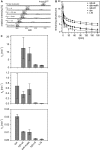
D/H-exchange kinetics in isotropic solution. Exhaustively deuterated peptides were assayed for back-exchange. (A) Exemplary mass spectra of the triply-charged syb-wt ion at different time points. The spectrum at t = 0 min was recorded after exchange under stop conditions (3 min incubation on ice at pH 2.5) where only very labile deuteriums exchange (see text for details). Low intensity isotopic envelopes at calculated masses ∼22 Da above the dominant envelopes likely originate from Na+-adducts. (B) A comparison of the exchange kinetics exhibited by syx, syb-wt, syb-multA, syb-L8, and L16. The data points at t = 0 correspond to the numbers of amide deuteriums seen after exchange under stop conditions. The data were fit with a three-term exponential function assuming D = 19 at t = 0 min (continuous lines; see Experimental Procedures for details). (C) Comparison of exchange rate constants that were calculated for deuterium classes whose size was equivalent for each peptide. No kA value syx is given for syx, whose kA is at least as high as that of syb but could not be precisely calculated due to improper curve fitting at the earliest time points. All values represent means ± SD of at least three independent measurements.
FIGURE 4
FIGURE 4
Peak-width analysis. Peak widths at 50% of maximal peak height were determined from the deconvoluted mass spectra obtained in 60% (v/v) TFE, 10 mM NH4Ac, pH 7.4 at 20°C and plotted as a function of reaction time from t = 0.5 min to t = 3 h. Representative spectra were used in all cases except for syx, where lower signal/noise ratios resulted in a greater apparent variability of peak widths; syx peak widths were thus averaged from three independent spectra.
FIGURE 5
FIGURE 5
H/D-exchange kinetics in liposomal membranes. Peptides were reconstituted into liposomal membranes made of DMPC at P/L = 0.01 and assayed for their exchange kinetics. The data points at t = 0 correspond to the numbers of amide deuteriums seen after exchange for 3 min under stop conditions. The data were fit with a three-term exponential function (continuous lines; see Experimental Procedures for details). All values represent means ± SD of at least three independent measurements.

Similar articles

Cited by

References

    1. Farrens, D. L., C. Altenbach, K. Yang, W. L. Hubbell, and H. G. Khorana. 1996. Requirement of rigid-body motion of transmembrane helices for light activation of rhodopsin. Science. 274:768–770. - PubMed
    1. Klingenberg, M. 2005. Ligand-protein interaction in biomembrane carriers. The induced transition fit of transport catalysis. Biochemistry. 44:8563–8570. - PubMed
    1. Matthews, E. E., M. Zoonens, and D. M. Engelman. 2006. Dynamic helix interactions in transmembrane signaling. Cell. 127:447–450. - PubMed
    1. Perozo, E., D. M. Cortes, P. Sompornpisut, A. Kloda, and B. Martinac. 2002. Open channel structure of MscL and the gating mechanism of mechanosensitive channels. Nature. 418:942–948. - PubMed
    1. Cordes, F. S., J. N. Bright, and M. S. Sansom. 2002. Proline-induced distortions of transmembrane helices. J. Mol. Biol. 323:951–960. - PubMed

Publication types

LinkOut - more resources