. 2008 Aug;95(4):1928-44.
doi: 10.1529/biophysj.107.126995. Epub 2008 May 2.

Biophysical characterization of the unstructured cytoplasmic domain of the human neuronal adhesion protein neuroligin 3

Affiliations

Biophysical characterization of the unstructured cytoplasmic domain of the human neuronal adhesion protein neuroligin 3

Aviv Paz et al. Biophys J. 2008 Aug.

Abstract

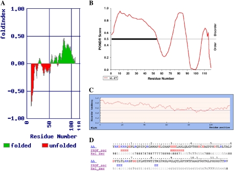
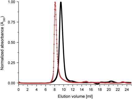
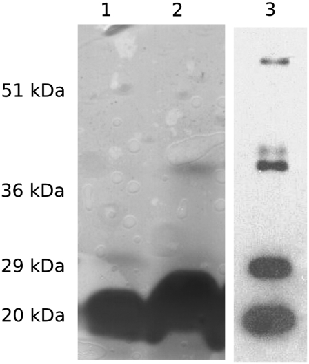
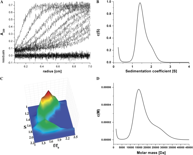
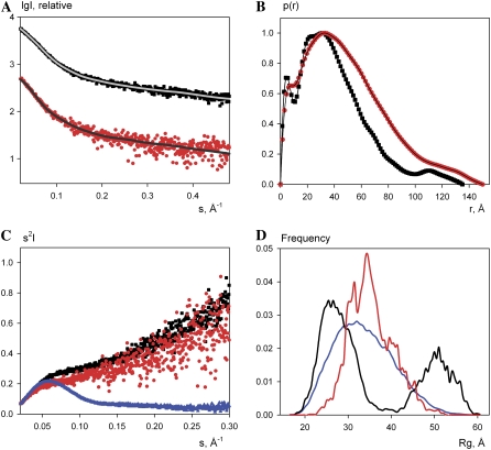
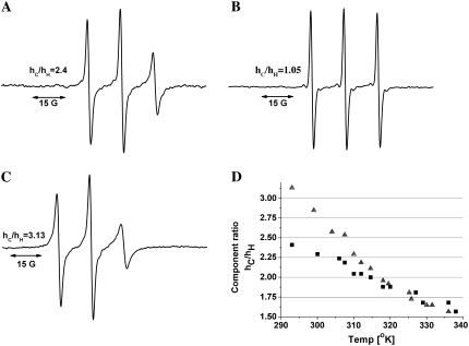
Cholinesterase-like adhesion molecules (CLAMs) are a family of neuronal cell adhesion molecules with important roles in synaptogenesis, and in maintaining structural and functional integrity of the nervous system. Our earlier study on the cytoplasmic domain of one of these CLAMs, the Drosophila protein, gliotactin, showed that it is intrinsically unstructured in vitro. Bioinformatic analysis suggested that the cytoplasmic domains of other CLAMs are also intrinsically unstructured, even though they bear no sequence homology to each other or to any known protein. In this study, we overexpress and purify the cytoplasmic domain of human neuroligin 3, notwithstanding its high sensitivity to the Escherichia coli endogenous proteases that cause its rapid degradation. Using bioinformatic analysis, sensitivity to proteases, size exclusion chromatography, fluorescence correlation spectroscopy, analytical ultracentrifugation, small angle x-ray scattering, circular dichroism, electron spin resonance, and nuclear magnetic resonance, we show that the cytoplasmic domain of human neuroligin 3 is intrinsically unstructured. However, several of these techniques indicate that it is not fully extended, but becomes significantly more extended under denaturing conditions.


Figures

FIGURE 1
FIGURE 1
Domain architecture of the CLAMs. All CLAMs contain a ChE-domain (shaded), which localizes to the extracellular compartment either as a secreted protein or as the extracellular domain of a single-pass transmembrane protein. Both Gli and NL are type I membrane proteins that bear a C-terminal PDZ recognition motif, while Nrt is a type II membrane protein. The percentage identity to human AChE is shown in each extracellular domain. Dm stands for Drosophila melanogaster and h stands for human. The hAChE scale bar represents 100 amino acids.
FIGURE 2
FIGURE 2
Bioinformatics analysis of hNL3-cyt. (A) Foldindex prediction; (B) PONDR prediction. Both algorithms predict that the 50 N-terminal amino acids of hNL3-cyt are disordered; (C) IUPred prediction for hNL3-cyt; and (D) PROF secondary structure prediction (in the PROF_sec line, H denotes helix and E denotes strand). Rel_sec line is the reliability index, ranging from 0 (low) to 9 (high).
FIGURE 3
FIGURE 3
SDS-PAGE of partially purified hNL3-cyt extracted under denaturing or nondenaturing conditions. hNL3-cyt was extracted from the bacterial pellet either under denaturing conditions (8 M urea/100 mM NaH2PO4/10 mM Tris, pH 8.0) or nondenaturing conditions (500 mM NaCl/50 mM NaH2PO4/10 mM imidazole, pH 8.0, containing a broad-range protease inhibitor cocktail). In both cases, extraction was at 4°C. Both extracts underwent an initial purification step which involved absorption on a Ni-column followed by elution using a pH gradient for the protein extracted under denaturing conditions, and an imidazole gradient for the sample extracted under nondenaturing conditions (for details, see Materials and Methods). Lane 1, molecular weight markers; Lane 2, hNL3-cyt purified after extraction under denaturing conditions; and Lane 3, hNL3-cyt purified after extraction under nondenaturing conditions. Western blotting, using anti-His antibodies, reveals that many of the fast-moving species below the band of intact hNL3-cyt (arrow) are degradation products (not shown).
FIGURE 4
FIGURE 4
Digestion of hNL3-cyt and TcAChE by proteinase K. Digestion conditions were as described under Materials and Methods. (A and B) SDS-PAGE of AChE (A) and hNL3-cyt (B) after digestion by proteinase K. Times of digestion, in minutes, are marked above each lane.
FIGURE 5
FIGURE 5
Size exclusion chromatography of hNL3-cyt. hNL3-cyt eluted at 8.1 ml under denaturing conditions (red), and at 9.4 ml under nondenaturing conditions (black). A perfect sphere with the molecular weight of hNL3-cyt would be predicted to elute at 11.85 ml.
FIGURE 6
FIGURE 6
Fluorescence correlation curves for ATTO-488 labeled hNL3-cyt. Measurements in 0.01% Tween/50 mM NaCl/50 mM sodium phosphate, pH 7.0 (▪). Measurements in 0.01% Tween/6M Gdn.HCl/12.5 mM NaCl/12.5 mM sodium phosphate, pH 7.0 (•). The green solid lines represent fits of the data to the model in Eq. 2. Residuals of data fits are displayed at the bottom of the figure for both the two correlation curves and for the fits.
FIGURE 7
FIGURE 7
SDS-PAGE and Western-blot of hNL3-cyt after nondenaturing size exclusion chromatography. hNL3-cyt elutes in several oligomeric states, which are stable under the conditions of SDS-PAGE. Lanes 1 and 2, SDS-PAGE of hNL3-cyt loaded at 2.4 mg/ml, and 4.8 mg/ml; lane 3, Western-blot of hNL3-cyt using anti-His antibody, which displays at least three distinct oligomeric states.
FIGURE 8
FIGURE 8
Analytical ultracentrifugation of hNL3-cyt. An AUC sedimentation velocity experiment was run at 0.3 mg/ml in PBS adjusted to pH 8.0. (A) Sedimentation data (solid line) and c(s) analysis fit (dotted line) of every 10th scan. Residuals are shown at the bottom, with a root mean-square deviation of 0.022. (B) c(s) distribution of hNL3-cyt. hNL3-cyt has a broad distribution with a principal peak (S = 1.33), and a shoulder (∼2 S). (C) Plot of S versus the frictional ratio, f/fo, derived from the c(s,fr) analysis. Both populations of hNL3-cyt have high f/fo values, indicating that both are extended in solution. (D) c(M) distribution of the same data. The first peak has a maximum at 15,227 ± 300 Da, in excellent agreement with the molecular weight predicted from the sequence.
FIGURE 9
FIGURE 9
SAXS measurements on hNL3-cyt. (A) X-ray scattering patterns of hNL3-cyt under denaturing (•) and nondenaturing conditions (▪). The fits of the ensembles selected by EOM are displayed as gray lines. (B) Distance distribution functions of the measurements shown in panel A. (C) Kratky plots (red and black traces) derived from the data in panel A and of the globular protein, BSA (blue trace), which served as a folded control. (D) Radii of gyration distributions of hNL3-cyt under denaturing (red line) and nondenaturing conditions (black line) and comparison with a pool of randomly generated structures (blue line).
FIGURE 10
FIGURE 10
CD spectra of hNL3-cyt. All spectra were collected at an hNL3-cyt concentration of 12 μM with the appropriate buffer corrections. (A) CD spectra of hNL3-cyt in 10 mM potassium phosphate (KPi), pH 8.0, at various temperatures (solid line, 5°C; dash-dotted line, 25°C; and dotted line, 45°C). (B) Effect of 7M Gdn.HCl and temperature on the CD spectrum of hNL3-cyt (solid line, 10 mM KPi, pH 8.0, at 5°C; long-dash, short-dash line, −7 M Gdn.HCl at 5°C; dash-dotted line, 7 M Gdn.HCl at 25°C; and dotted line, 7 M Gdn.HCl at 45°C). (C) Effect of 50% TFE and temperature on the CD spectrum of hNL3-cyt (solid line, 10 mM KPi, pH 8.0, at 5°C; long-dash, short-dash line, 50% TFE at 5°C; dotted line, 50% TFE at 25°C; and dash-dotted line, 50% TFE at 45°C). (D) Effect of 0.5 mM SDS and 1 mM DOPS liposomes on the CD spectrum of hNL3-cyt (solid line, 10 mM KPi, pH 8.0, 25°C; long-dash, short-dash line, 0.5 mM SDS; and dotted line, 1 mM DOPS).
FIGURE 11
FIGURE 11
ESR measurements on spin-labeled hNL3-cyt. (A) The ESR spectrum of spin-labeled hNlg3-cyt (in 150 mM NaCl/10 mM sodium phosphate, pH 7.0, at 20°C), reveals relatively sharp peaks that indicate rather limited restriction of the probe's mobility. (B) Disappearance of the relatively slow components, with concomitant appearance of a sharp triplet, when a 100-fold molar excess of DTT was added to the spin-labeled protein. (C) ESR spectrum obtained in the presence of 66% TFE, at 20°C. The peak intensity of the high-field component (hH) in the conjugate (which is very sensitive to label movement) decreases relative to that seen in the absence of TFE, suggesting a decrease in segmental mobility of the polypeptide. (D) Temperature-dependence of the (hC)/(hH) ratio. Both in the absence (▪) and presence of TFE (▴), the ratio decreases with temperature, reflecting an increase in mobility.
FIGURE 12
FIGURE 12
NMR measurements on hNL3-cyt. Contour plots of two-dimensional 15N{1H} NOE NMR spectra recorded without (A) and with (B) proton saturation. Spectra are shown at the same contour level. Contour levels in spectrum A are positive, and in panel B are negative. Arg NE denotes side-chain Nɛ-Hɛ correlations that are aliased in the 15N dimension from ∼84.7 ppm. One-dimensional traces taken from panels A and B at 120 ppm (15N) are shown in panels C and D, respectively.

Similar articles

Cited by

References

    1. Botti, S. A., C. E. Felder, J. L. Sussman, and I. Silman. 1998. Electrotactins: a class of adhesion proteins with conserved electrostatic and structural motifs. Protein Eng. 11:415–420. - PubMed
    1. Gilbert, M. M., and V. J. Auld. 2005. Evolution of clams (cholinesterase-like adhesion molecules): structure and function during development. Front. Biosci. 10:2177–2192. - PubMed
    1. Ichtchenko, K., Y. Hata, T. Nguyen, B. Ullrich, M. Missler, C. Moomaw, and T. C. Sudhof. 1995. Neuroligin 1: a splice site-specific ligand for β-neurexins. Cell. 81:435–443. - PubMed
    1. Gilbert, M., J. Smith, A. J. Roskams, and V. J. Auld. 2001. Neuroligin 3 is a vertebrate gliotactin expressed in the olfactory ensheathing glia, a growth-promoting class of macroglia. Glia. 34:151–164. - PubMed
    1. Bolliger, M. F., K. Frei, K. H. Winterhalter, and S. M. Gloor. 2001. Identification of a novel neuroligin in humans which binds to PSD-95 and has a widespread expression. Biochem. J. 356:581–588. - PMC - PubMed

Publication types

LinkOut - more resources