. 2008 Jul;14(7):1424-32.
doi: 10.1261/rna.755908. Epub 2008 May 2.

Extracellular plasma RNA from colon cancer patients is confined in a vesicle-like structure and is mRNA-enriched

Affiliations

Extracellular plasma RNA from colon cancer patients is confined in a vesicle-like structure and is mRNA-enriched

José Miguel García et al. RNA. 2008 Jul.

Abstract

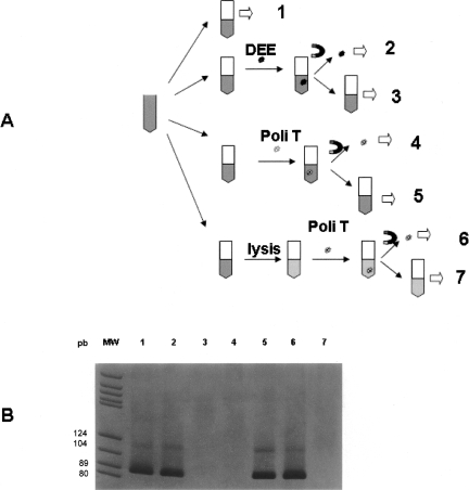
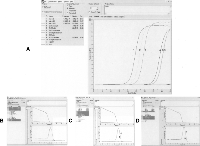
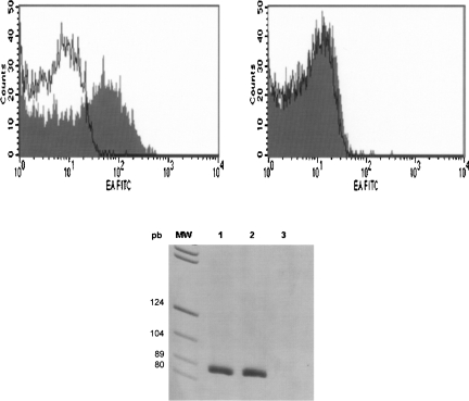
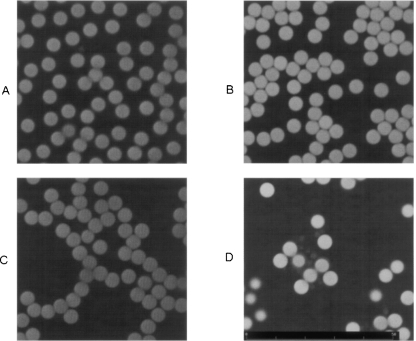
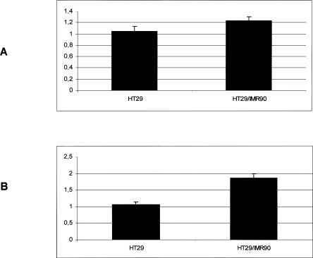
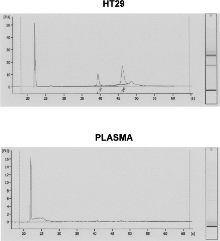
Little is yet known about the origin and protective mechanism of free nucleic acids in plasma. We investigated the possibility of these free nucleic acids being particle associated. Plasma samples from colon cancer patients and cell culture media were subjected to various antibody incubations, ultracentrifugation, and RNA extraction protocols for total RNA, epithelial RNA, and mRNA. Flow cytometry using a Ber-EP4 antibody and confocal laser microscopy after staining with propidium iodide were also performed. mRNA levels of the LISCH7 and SDHA genes were determined in cells and in culture media. Ber-EP4 antibody and polystyrene beads coated with oligo dT sequences were employed. We observed that, after incubation, total RNA and mRNA were always detected after membrane digestion, and that epithelial RNA was detected before this procedure. In ultracentrifugation, mRNA was caught in the supernatant only if a former lysis mediated or in the pellet if there was no previous digestion. Flow cytometry determinations showed that antibody-coated microbeads keep acellular structures bearing epithelial antigens apart. Confocal laser microscopy made 1- to 2-microm-diameter particles perceptible in the vicinity of magnetic polystyrene beads. Relevant differences were observed between mRNA of cells and culture media, as there was a considerable difference in LISCH7 mRNA levels between HT29 and IMR90 cell co-cultures and their culture media. Our results support the view that extracellular RNA found in plasma from cancer patients circulates in association with or is protected in a multiparticle complex, and that an active release mechanism by tumor cells may be a possible origin.


Figures

FIGURE 1.
FIGURE 1.
(A) Representative outline of the incubation processes of different aliquots from a plasma sample. Step 1, Total RNA extraction; step 2, epithelial RNA extraction after incubation with DEE; step 3, total RNA extraction after withdrawal of DEE microspheres; steps 4 and 6, mRNA extraction after incubation with the Dynabeads mRNA DIRECT kit; steps 5 and 7, total RNA extraction after withdrawal of the oligo dT sequence-coated microspheres. Steps 6 and 7, unlike steps 4 and 5, were carried out after lysis of the plasma sample. (B) Electrophoresis of the SDHA amplification products in each of the previous incubation processes. Each number corresponds to the same number in panel A. DEE, Dynabeads Epithelial Enrich (Epithelial Antigen-coated polystyrene beads); Poli T, polystyrene beads coated with oligo dT sequence; MW, molecular weight marker; magnet symbol, magnetic isolation.
FIGURE 2.
FIGURE 2.
SDHA real-time PCR results from the ultracentrifugation assay. “Pellet” represents mRNA extraction from the pellet after ultracentrifugation; “Supernatant” represents mRNA extraction from the supernatant after ultracentrifugation; and “Epithelial Enrich” represents epithelial RNA extraction from the supernatant after ultracentrifugation. Before ultracentrifugation, one of the samples was subjected to lysis (SD, sample digested), whereas the other was not (SND, sample non-digested). (A) Fluorescence acquisition. (B) Melting curves from serial dilution of cDNA (13) and positive sample (4). (C) Melting curves from positive sample (4) and non-digested samples (5). (D) Melting curves from the positive sample (4) and digested samples (9). Retro-transcription of an H2O sample (H2O RT), or an H2O sample was used directly as a negative control (H2O).
FIGURE 3.
FIGURE 3.
(Left panel) FACS analyses of plasma incubated with monoclonal antibody mouse anti-human epithelial antigen, clone Ber-EP4, filled trace, prior to DEE incubation or after incubation and withdrawal DEE (right panel). A mouse IgG1-FITC irrelevant Ab was used as a negative control of staining in both cases (open trace). (Bottom panel) SDHA real-time PCR results from RNA extracted from plasma before incubation with DEE (lane 1), DEE fraction (lane 2), and plasma after incubation and withdrawal DEE (lane 3).
FIGURE 4.
FIGURE 4.
Result of confocal laser examination of different samples incubated in the presence of super-paramagnetic polystyrene beads coated with Ber-EP4 Ab. (A) PBS without staining with propidium iodide (PI). (B) The same sample stained with PI. (C) Plasma from a colon cancer patient without PI staining. (D) The same sample stained with PI, showing small masses in the vicinity of the microbeads.
FIGURE 5.
FIGURE 5.
LISCH7 mRNA levels in HT29 cells growing alone or in the presence of IMR90 fibroblasts (A), 14.7% increase; or their respective culture media (B), 74.67% increase. Standardized mean values and standard deviations obtained in four independent experiments are shown.
FIGURE 6.
FIGURE 6.
(Top panel) RNA from the HT29 cell line and from plasma from colon cancer patients, detected using a bioanalyzer. (Bottom panel) No or very low ribosomal RNA levels are present in the RNA from the plasma sample, as well as more small RNAs than in the RNA obtained from HT29 cell line.

Similar articles

Cited by

References

    1. Anker P., Stroun M., Maurice P.A. Spontaneous release of DNA by human blood lymphoctyes as shown in an in vitro system. Cancer Res. 1975;35:2375–2382. - PubMed
    1. Anker P., Lefort F., Vasioukhin V., Lyautey J., Lederrey C., Chen X.Q., Stroun M., Mulcahy H.E., Farthing M.J. K-ras mutations are found in DNA extracted from the plasma of patients with colorectal cancer. Gastroenterology. 1997;112:1114–1120. - PubMed
    1. Anker P., Mulcahy H., Chen X.Q., Stroun M. Detection of circulating tumour DNA in the blood (plasma/serum) of cancer patients. Cancer Metastasis Rev. 1999;18:65–73. - PubMed
    1. Bhowmick N.A., Chytil A., Plieth D., Gorska A.E., Dumont N., Shappell S., Washington M.K., Neilson E.G., Moses H.L. TGF-β signaling in fibroblasts modulates the oncogenic potential of adjacent epithelia. Science. 2004a;303:848–851. - PubMed
    1. Bhowmick N.A., Neilson E.G., Moses H.L. Stromal fibroblasts in cancer initiation and progression. Nature. 2004b;432:332–337. - PMC - PubMed

Publication types

LinkOut - more resources