Comparative Study
. 2008 May;211(Pt 10):1594-602.
doi: 10.1242/jeb.017244.

Synergy and specificity of two Na+-aromatic amino acid symporters in the model alimentary canal of mosquito larvae

Affiliations
Comparative Study

Synergy and specificity of two Na+-aromatic amino acid symporters in the model alimentary canal of mosquito larvae

Bernard A Okech et al. J Exp Biol. 2008 May.

Abstract

The nutrient amino acid transporter (NAT) subfamily is the largest subdivision of the sodium neurotransmitter symporter family (SNF; also known as SLC6; HUGO). There are seven members of the NAT population in the African malaria mosquito Anopheles gambiae, two of which, AgNAT6 and AgNAT8, preferably transport indole- and phenyl-branched substrates, respectively. The relative expression and distribution of these aromatic NATs were examined with transporter-specific antibodies in Xenopus oocytes and mosquito larval alimentary canal, representing heterologous and tissue expression systems, respectively. NAT-specific aromatic-substrate-induced currents strongly corresponded with specific accumulation of both transporters in the plasma membrane of oocytes. Immunolabeling revealed elevated expressions of both transporters in specific regions of the larval alimentary canal, including salivary glands, cardia, gastric caeca, posterior midgut and Malpighian tubules. Differences in relative expression densities and spatial distribution of the transporters were prominent in virtually all of these regions, suggesting unique profiles of the aromatic amino acid absorption. For the first time reversal of the location of a transporter between apical and basal membranes was identified in posterior and anterior epithelial domains corresponding with secretory and absorptive epithelial functions, respectively. Both aromatic NATs formed putative homodimers in the larval gut whereas functional monomers were over-expressed heterologously in Xenopus oocytes. The results unequivocally suggest functional synergy between substrate-specific AgNAT6 and AgNAT8 in intracellular absorption of aromatic amino acids. More broadly, they suggest that the specific selectivity, regional expression and polarized membrane docking of NATs represent key adaptive traits shaping functional patterns of essential amino acid absorption in the metazoan alimentary canal and other tissues.


Figures

Fig. 1
Fig. 1
Western blot of AgNATs expression in Xenopus eggs and larval midgut tissues. Western blot analysis of AgNAT6 and AgNAT8 expression are shown in left and right panels, respectively. Lanes: ps, pre-immune sera; ab, purified antibodies; dw, tr and ts, membrane fractions from deionized water- or transcript-injected oocytes and midgut tissue, respectively. Numbers are protein mass references (in kDa). Arrowheads indicate putative monomer and homodimer bands. Asterisks indicate dimers in the tissue fractions.
Fig. 2
Fig. 2
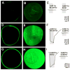
Functional expression of AgNAT6 and AgNAT8 in Xenopus oocytes. Images represent results of immunolabeling of oocytes with transporter-specific antibodies after oocytes were injected with (A,B) deionized water or (D–H) specific aromatic NAT transcripts, i.e. AgNAT6 (D,E) and AgNAT8 (G,H). Fourth day post-injection oocytes are shown. Control oocytes treated with AgNAT8 antibodies (A,B) and AgNAT6 antibodies (data not shown) produce very low background labeling. Optical mid-oocyte sections (A,D,G) and surface reconstruction from multiple confocal frames (B,E,H) are shown along with amino acid-induced currents (C,F,I).
Fig. 3
Fig. 3
Relative transport efficiency of aromatic NATs. Normalized means (bars) of selected substrate-induced currents were acquired from independent oocytes (N ≥ 3 for each data point). All responses were measured using standard conditions (98 mmol·l−1 Na+ oocyte perfusion saline, 50 mV holding potential, and 1 mmol·l−1 of organic substrate concentrations). Data sets for AgNAT6 (blue set) and AgNAT8 (red set) were normalized relative to Trp- and Phe-induced responses and sorted with respect to amplitudes of induced currents and chemical substrate properties. Final data sets were fitted using a four parameters Gaussian pick function: f=y0+aexp{−0.5[(xx0)/b]^2} indicated by red and blue lines. Substrate groups are indicated by different font styles: neurotransmitter, underlined; aromatic substrates, solid black; and all others gray.
Fig. 4
Fig. 4
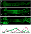
Relative distribution of AgNAT6 and AgNAT8 in the larval alimentary canal. Isolated alimentary canal of fourth instar larvae were labeled with (A) preimmune serum, (B) AgNAT6- and (C) AgNAT8-specific antibodies. Primary antibody binding areas were visualized using Alexa Fluor 467 secondary antibodies, producing green fluorescent signals. CA, cardia; GC, gastric caeca; AMG, anterior midgut; PMG, posterior midgut; MT, Malpighian tubes; RG, rectal gland. (D) Relative fluorescence intensities are shown along the larval alimentary canal (scans from A,B,C are represented by black, green and magenta lines, respectively). Rectangular area scan signal values are indicated by dotted lines; scans filtered by running average values indicated by solid lines. Scale bar, 1 mm.
Fig. 5
Fig. 5
Immunolabeling of AgNATs on frozen sections of the larval alimentary canal. Immunolabeling of frozen sections with AgNAT6 (left site of the panel) AgNAT8 (right site of the panel) epitope-specific purified antibodies (green channel), along with actin (TRITC-phalloidin, red channel)- and nuclei (DRAQ-5, blue channel)-specific labeling are shown. The actin and nuclei channels were turn off on a few sections to improve overall visualization. The red channel in general represents actin of the muscular envelop around the gut, and corresponds to the position of the basal membrane, except in the salivary gland and Malpighian tubules where it indicates the actin of microvillae and the apical membrane, respectively. The position of the apical membrane is indicated by white arrows. Approximate positions of individual sections are shown on the central diagram. SG, salivary gland; CA, cardia; GC, gastric caeca; AMG, anterior midgut; PMG, posterior midgut; MT, Malpighian tubes. Control sections of the AMG (control AMG) and PMG (control PMG) incubated with pre-bleed serum are shown in the bottom left corner. Scale bars, 50 μm.
Fig. 6
Fig. 6
A diagram of the relative distribution of aromatic AgNATs in the model system of the larval alimentary canal. A summary diagram composed from whole-mount and frozen section preparations is shown (N>20 for each transporter). Green dotted lines indicate membrane docking of the transporters. Colored arrows indicate direction of transport at specified locations. Their colors show either the same (green), specific (black) and opposed directions of substrate transport by AgNAT6 (top part) compared with AgNAT8 (bottom part). Gray arrows show a putative circuit of cation recycling via alkalinization (pink gradient region) and NAT-coupled pathway. Empty arrow indicates location of a putative H+ V-ATPase and cation exchanger-coupled mechanism for cation translocation in the AMG. SG, salivary gland; GC, gastric caeca; AMG, anterior midgut; PMG, posterior midgut; MT, Malpighian tubes; RG, rectal gland; cm, caecal membrane; pm, peritrophic membrane.

Similar articles

Cited by

References

    1. Assis P, Boudko DY, Meleshkevitch EA, Phung E. Molecular expression and electrochemical analysis of phenylalanine-tyrosine transporter from Anopheles gambiae larvae. FASEB J. 2004;18:A1269.
    1. Bohmer C, Broer A, Munzinger M, Kowalczuk S, Rasko JEJ, Lang F, Broer S. Characterization of mouse amino acid transporter B(0)AT1 (slc6a19) Biochem J. 2005;389:745–751. - PMC - PubMed
    1. Bossi E, Soragna A, Miszner A, Giovannardi S, Frangione V, Peres A. Oligomeric structure of the neutral amino acid transporters KAAT1 and CAATCH1. Am J Physiol. 2007;292:C1379–C1387. - PubMed
    1. Boudko DY, Moroz LL, Harvey WR, Linser PJ. Alkalinization by chloride/bicarbonate pathway in larval mosquito midgut. Proc Natl Acad Sci USA. 2001a;98:15354–15359. - PMC - PubMed
    1. Boudko DY, Moroz LL, Linser PJ, Trimarchi JR, Smith PJS, Harvey WR. In situ analysis of pH gradients in mosquito larvae using noninvasive, self-referencing, pH-sensitive microelectrodes. J Exp Biol. 2001b;204:691–699. - PubMed

Publication types