

Massive hematuria due to a congenital renal arteriovenous malformation mimicking a renal pelvis tumor: a case report
- PMID: 18457585
- PMCID: PMC2390576
- DOI: 10.1186/1752-1947-2-144
Massive hematuria due to a congenital renal arteriovenous malformation mimicking a renal pelvis tumor: a case report
Abstract
Introduction: Congenital renal arteriovenous malformations (AVMs) are very rare benign lesions. They are more common in women and rarely manifest in elderly people. In some cases they present with massive hematuria. Contemporary treatment consists of transcatheter selective arterial embolization which leads to resolution of the hematuria whilst preserving renal parenchyma.
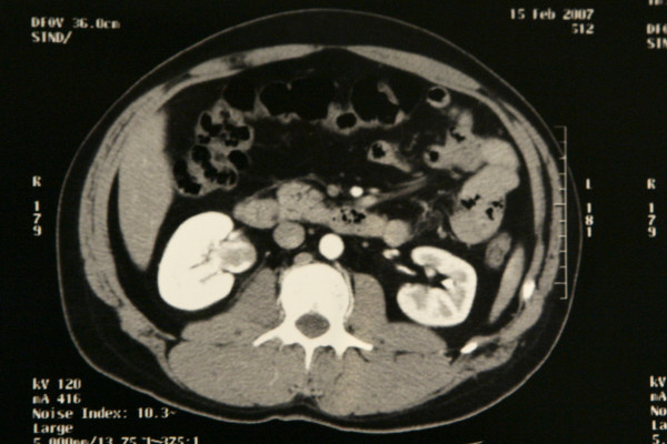
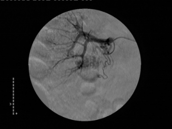
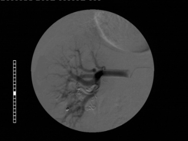
Case presentation: A 72-year-old man, who was heavy smoker, presented with massive hematuria and flank pain. CT scan revealed a filling defect caused by a soft tissue mass in the renal pelvis, which initially led to the suspicion of a transitional cell carcinoma (TCC) of the upper tract, in view of the patient's age and smoking habits. However a subsequent retrograde study could not depict any filling defect in the renal pelvis. Selective right renal arteriography confirmed the presence of a renal AVM by demonstrating abnormal arterial communication with a vein with early visualization of the venous system. At the same time successful selective transcatheter embolization of the lesion was performed.
Conclusion: This case highlights the importance of careful diagnostic work-up in the evaluation of upper tract hematuria. In the case presented, a congenital renal AVM proved to be the cause of massive upper tract hematuria and flank pain in spite of the initial evidence indicating the likely diagnosis of a renal pelvis tumor.
Figures
References
-
- Rosen RJ, Ryles TS. Arterial venous malformations. In: Strandness DE, Van Breda A, editor. Vascular disease Surgical and Interventional Therapy. Vol. 2. New York, Churchill Livingstone; 1994. pp. 1121–37.
-
- Liu W, Esler SJ, Kenny BJ. Low-dose nonenhanced helical CT of renal colic: assessment of ureteric stone detection and measurement of effective dose equivalent. Radiology. 2000;215:51–54. - PubMed
-
- Saito S, Iigaya T, Koyama Y. Transcatheter embolization for the rupture of congenital arteriovenous malformation of the kidney in pregnancy. J Urol. 1987;137:964–5. - PubMed
LinkOut - more resources
Full Text Sources
