

Evidence of Brucella abortus OPS dictating uptake and restricting NF-kappaB activation in murine macrophages
- PMID: 18457975
- PMCID: PMC2752336
- DOI: 10.1016/j.micinf.2008.01.005
Evidence of Brucella abortus OPS dictating uptake and restricting NF-kappaB activation in murine macrophages
Abstract
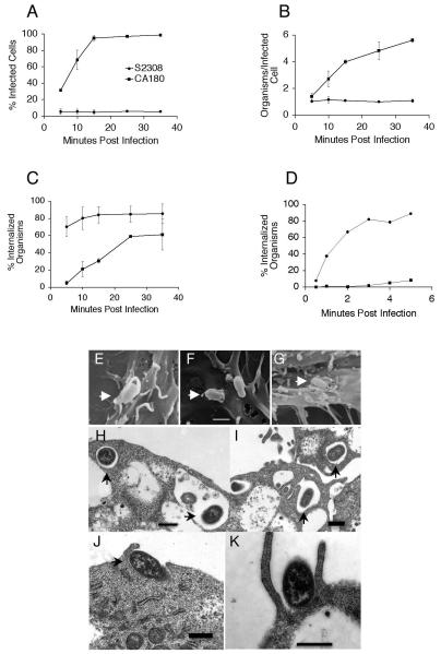
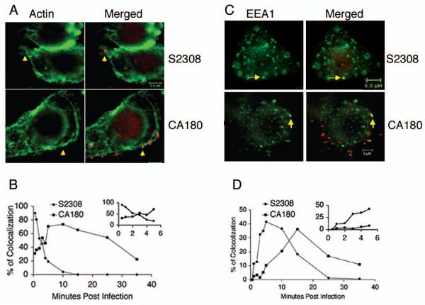
Smooth Brucella abortus S2308 is virulent while rough derivatives are attenuated. Intracellular killing is often blamed for these differences. In the studies described, uptake kinetics and interaction of S2308 and S2308 manBA::Tn5 (CA180) rough mutants with macrophages were investigated. The results revealed that smooth B. abortus was rapidly internalized, achieving a maximum level in less than 5 min without additional uptake over the next 30 min. In contrast, continued uptake of the rough mutant was observed and only achieves a maximum level after 30 min. The results were confirmed by the differences in F-actin polymerization, lipid raft staining, early endosome colocalization and electron microscopic observations after smooth and rough Brucella infection. We also demonstrated for the first time that uptake of S2308, but not rough mutant CA180 was PI3-kinase and toll-like receptor 4 (TLR4) dependent. Differences in uptake were associated with differences in macrophage activation with regard to NF-kappaB translocation and cytokine production. These results provide evidence that the presence of B. abortus OPS dictates the interactions between Brucella and specific cell surface receptors minimizing macrophage activation and enhancing Brucella survival and/or persistence.
Figures


Similar articles
-
Inflammatory response of TLR4 deficient spleen macrophages (CRL 2471) to Brucella abortus S19 and an isogenic ΔmglA deletion mutant.Int J Med Microbiol. 2016 May;306(3):141-51. doi: 10.1016/j.ijmm.2016.02.006. Epub 2016 Feb 20. Int J Med Microbiol. 2016. PMID: 26946956
-
Role of Toll-like receptor 4 in induction of cell-mediated immunity and resistance to Brucella abortus infection in mice.Infect Immun. 2004 Jan;72(1):176-86. doi: 10.1128/IAI.72.1.176-186.2004. Infect Immun. 2004. PMID: 14688095 Free PMC article.
-
Brucella abortus rough mutants induce macrophage oncosis that requires bacterial protein synthesis and direct interaction with the macrophage.Infect Immun. 2006 May;74(5):2667-75. doi: 10.1128/IAI.74.5.2667-2675.2006. Infect Immun. 2006. PMID: 16622203 Free PMC article.
-
TLR2 and TLR4 signaling pathways are required for recombinant Brucella abortus BCSP31-induced cytokine production, functional upregulation of mouse macrophages, and the Th1 immune response in vivo and in vitro.Cell Mol Immunol. 2014 Sep;11(5):477-94. doi: 10.1038/cmi.2014.28. Epub 2014 Apr 28. Cell Mol Immunol. 2014. PMID: 24769793 Free PMC article.
-
The role of innate immune signals in immunity to Brucella abortus.Front Cell Infect Microbiol. 2012 Oct 25;2:130. doi: 10.3389/fcimb.2012.00130. eCollection 2012. Front Cell Infect Microbiol. 2012. PMID: 23112959 Free PMC article. Review.
Cited by
-
Toll-like receptors are critical for clearance of Brucella and play different roles in development of adaptive immunity following aerosol challenge in mice.Front Cell Infect Microbiol. 2012 Sep 7;2:115. doi: 10.3389/fcimb.2012.00115. eCollection 2012. Front Cell Infect Microbiol. 2012. PMID: 22973560 Free PMC article.
-
Key immunity characteristics of diverse stages of brucellosis in rural population from Inner Mongolia, China.Infect Dis Poverty. 2022 Jun 4;11(1):63. doi: 10.1186/s40249-022-00989-7. Infect Dis Poverty. 2022. PMID: 35659087 Free PMC article.
-
Entry and elimination of marine mammal Brucella spp. by hooded seal (Cystophora cristata) alveolar macrophages in vitro.PLoS One. 2013 Jul 25;8(7):e70186. doi: 10.1371/journal.pone.0070186. Print 2013. PLoS One. 2013. PMID: 23936159 Free PMC article.
-
When the Going Gets Rough: The Significance of Brucella Lipopolysaccharide Phenotype in Host-Pathogen Interactions.Front Microbiol. 2021 Jul 15;12:713157. doi: 10.3389/fmicb.2021.713157. eCollection 2021. Front Microbiol. 2021. PMID: 34335551 Free PMC article. Review.
-
AutoCellANLS: An Automated Analysis System for Mycobacteria-Infected Cells Based on Unstained Micrograph.Biomolecules. 2022 Feb 1;12(2):240. doi: 10.3390/biom12020240. Biomolecules. 2022. PMID: 35204741 Free PMC article.
References
-
- Pizarro-Cerda J, Moreno E, Gorvel JP. Invasion and intracellular trafficking of Brucella abortus in nonphagocytic cells. Microbes Infect. 2000;2(7):829–35. - PubMed
-
- Ackermann MR, Cheville NF, Deyoe BL. Bovine ileal dome lymphoepithelial cells: endocytosis and transport of Brucella abortus strain 19. Vet. Pathol. 1988;25:28–35. - PubMed
-
- Guzman-Verri C, Chaves-Olarte C, von Eichel-Streiber C, Lopez-Goni I, Thelestam M, Arvidson S, Gorvel JP, Moreno E. GTPases of the Rho subfamily are required for Brucella abortus internalization in nonprofessional phagocytes: direct activation of Cdc42. J Biol Chem. 2001;276(48):44435–43. - PubMed
-
- Detilleux PG, Deyoe BL, Cheville NF. Effect of endocytic and metabolic inhibitors on the internalization and intracellular growth of Brucella abortus in Vero cells. Am J Vet Res. 1991;52(10):1658–64. - PubMed
Publication types
MeSH terms
Substances
Grants and funding
LinkOut - more resources
Full Text Sources
