. 2008 Jul;28(13):4261-74.
doi: 10.1128/MCB.02252-07. Epub 2008 May 5.

GTPase-mediated regulation of the unfolded protein response in Caenorhabditis elegans is dependent on the AAA+ ATPase CDC-48

Affiliations

GTPase-mediated regulation of the unfolded protein response in Caenorhabditis elegans is dependent on the AAA+ ATPase CDC-48

Marie-Elaine Caruso et al. Mol Cell Biol. 2008 Jul.

Abstract

When endoplasmic reticulum (ER) homeostasis is perturbed, an adaptive mechanism is triggered and named the unfolded protein response (UPR). Thus far, three known UPR signaling branches (IRE-1, PERK, and ATF-6) mediate the reestablishment of ER functions but can also lead to apoptosis if ER stress is not alleviated. However, the understanding of the molecular mechanisms integrating the UPR to other ER functions, such as membrane traffic or endomembrane signaling, remains incomplete. We consequently sought to identify new regulators of UPR-dependent transcriptional mechanisms and focused on a family of proteins known to mediate, among other, ER-related functions: the small GTP-binding proteins of the RAS superfamily. To this end, we used transgenic UPR reporter Caenorhabditis elegans strains as a model to specifically silence small-GTPase expression. We show that the Rho subfamily member CRP-1 is an essential component of UPR-induced transcriptional events through its physical and genetic interactions with the AAA+ ATPase CDC-48. In addition, we describe a novel signaling module involving CRP-1 and CDC-48 which may directly link the UPR to DNA remodeling and transcription control.


Figures

FIG. 1.
FIG. 1.
Identification and characterization of powerful ER stress GFP reporter genes. (A) List of ER stress promoter::gfp reporter genes successfully constructed, with the strain name and the human ortholog information. (B) Fluorescence induction of the eight GFP reporter genes when treated with 5 μg/ml TM. Fluorescence level was quantified on entire transgenic worm populations (≥1,000 worms per conditions) using the Copas Biosort. The increase is the ratio between the average fluorescence under TM conditions compared to the basal fluorescence level of each strain. (C) Expression pattern of each GFP reporter strain in living animals. Intestine expression and neuronal expression are represented in light gray and dark gray, respectively. (D) Fluorescence microscopy pictures for the three strains with the highest increases in fluorescence (tag-320, hsp-4, and ckb-2 strains) under basal conditions or after TM treatment.
FIG. 2.
FIG. 2.
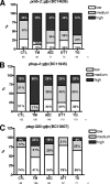
ER stressors TM, AZC, DTT, and TG act differentially on the three most responsive GFP reporter strains. Populations of (A) pckb-2::gfp (BC14636), (B) phsp-4::gfp (BC11945), and (C) ptag-320::gfp (BC13607) strains were treated with 5 μg/ml TM, 10 μM AZC, 2 mM DTT, and 5 μM TG for 5 hours and visualized and quantified using a fluorescence microscope. The percentage of worms with a low, medium, or high level of fluorescence was plotted against the different chemicals. Experiments were performed at least twice in duplicate. CTL, control.
FIG. 3.
FIG. 3.
Effect of crp-1, cdc-42, and sar-1 silencing on ckb-2 promoter activation upon ER stress. (A) Qualitative microscopic pictures for two controls (no RNAi induction [ctl] or GFP RNAi) and three GTPases that gave the most significant results for pckb-2::gfp reporter activation (encoded by crp-1, cdc-42, and sar-1). (B) Quantification of fluorescence induction of pckb-2::gfp subjected or not to crp-1 RNAi and exposed or not to 5 μg/ml TM.
FIG. 4.
FIG. 4.
CRP-1 interacts with a protein complex which includes CDC-48.1 and ATM-1. (A) SDS-PAGE of the GST-CRP-1 pull-down performed on precleared worm lysate, followed by mass spectrometry analysis (right). Peptides identified were annotated using the WormBase (http://www.wormbase.org/) sequence name as well as the Caenorhabditis Genetic Center (http://www.cbs.umn.edu/CGC/) three-letter name. (B, left) GST pull-down using GST-CRP-1 or GST-CDC-42 and mouse His6-p97/VCP/CDC-48 recombinant proteins expressed in bacteria and resolved on SDS-PAGE gel stained with Coomassie blue R250. (Right) His6-p97/VCP/CDC-48 pull-down was performed on wild-type (WT) (N2) or crp-1/ total worm extract followed by immunoblot analysis using anti (α)-CRP-1 antibodies (HyperOmics Farma, Inc.) or anti-p97 antibodies. (C) Transcriptional regulation of pckb-2::gfp reporter by cdc-48. pckb-2::gfp transgenic worms were subjected or not to cdc-48.1 RNAi as described in Materials and Methods. Following exposure to TM, pckb-2::gfp activation was visualized and quantified using fluorescence microscopy. (D) STRING network representation of the known and predicted interactions between proteins identified by mass spectrometry (white), VCP (p97/VCP/CDC-48) first-interacting proteins (gray), and ATM-1 first-interacting proteins (black). The edges are representative of the various interaction types available through STRING. Circles and squares are indicative of human and C. elegans genes, respectively (see also Table S3 in the supplemental material).
FIG. 5.
FIG. 5.
Physical interactions between CRP-1, HIM-6, and CDC-48. (A) Binary interactions between CRP-1 and either His6-HIM-6 or His6-CDC-48. The purified proteins were pulled-down using Ni-NTA-agarose beads, and the presence of GST-CRP-1 was revealed by immunoblotting (Ib) using anti-GST antibodies. (B) Binary interactions between His6-CDC-48.1 and Strep-tagged HIM-6 or His6-HIM-6 and Strep-tagged CDC-48.1 following purification of the complex by Ni-NTA-agarose beads and analysis by immunoblotting using anti-Strep tag antibodies. (C) Ternary complex including CRP-1, HIM-6, and CDC-48.1. Increasing concentrations of His6-CDC-48.1 were added to an equimolar mixture of GST-CRP-1 and Strep-tagged HIM-6. Following 1 h of incubation on ice, the complex was pulled-down using glutathione-Sepharose beads and the presence of HIM-6 in the complex was evaluated by immunoblotting using anti-Strep tag antibodies.
FIG. 6.
FIG. 6.
crp-1/, atm-1/, and cdc-48.1/ mutants are hypersensitive to TM and interact genetically. (A) Pictures showing the average sizes of wild-type (N2) and mutant worms when exposed to different concentrations of TM. (B) The percentage of progeny arrested in the L1 stage of development was plotted against the different concentrations of TM for the wild-type worms (N2) and cdc-48.1/ (tm544), crp-1/ (ok685), and atm-1/ (gk186) mutants. (C and D) The percentage of progeny arrested in L1 stages of development was plotted against the different concentrations of TM for cdc-48.1/ and atm-1/ mutants subjected of not to crp-1 RNAi (C) and for the atm-1/ mutant subjected of not to cdc-48 RNAi (D).
FIG. 7.
FIG. 7.
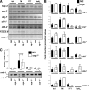
CRP-1-dependent regulation of the UPR transcriptional response. (A) Evaluation of the expression of the UPR target genes hsp-4, srp-7, dnj-7, cht-1, ckb-2, and F22E5.6 by RT-PCR analysis on mRNA isolated from worms treated or not with 5 μg/ml TM or 2 mM DTT for 5 h or 10 mM sodium azide for 90 min. PCR products are visualized following resolution by agarose gel electrophoresis, staining with ethidium bromide, and UV transillumination. Ctrl, control. (B) Quantification of the gels presented in panel A. Values relative to the expression of a control mRNA (ama-1) are reported and were obtained following normalization. The graphs represent the averages of three independent experiments ± standard deviations (SD). (C) Assessment of xbp-1 mRNA splicing using RT-PCR on mRNA collected as described above. PCR products are visualized followed resolution by agarose gel electrophoresis, staining with ethidium bromide, and UV transillumination (bottom). The graph shows the quantification of spliced xbp-1 mRNA (sXBP-1)/spliced plus unspliced xbp-1 mRNA (total XBP-1) as deduced from three independent experiments ± SD.
FIG. 8.
FIG. 8.
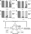
Genetic interaction map linking crp-1 and the UPR proximal sensors. (A to C) The percentage of progeny arrested in the L1 stage of development was plotted against different concentrations of TM (2 or 5 μg/ml) for N2 (A), pek-1/ (B), and atf-6/ (C) animals subjected or not to crp-1 RNAi. (D) The percentage of progeny arrested in the L1 stage of development was plotted against different concentrations of TM (2 or 5 μg/ml) for crp-1/ animals subjected to xbp-1 RNAi. (E) Schematic representation of CDC-48-dependent pathways proposed to explain the implication of CRP-1, HIM-6, and ATM-1 in the regulation of ER homeostasis during stress condition through DNA remodeling/transcriptional processes.

Similar articles

Cited by

References

    1. Ababou, M., S. Dutertre, Y. Lecluse, R. Onclercq, B. Chatton, and M. Amor-Gueret. 2000. ATM-dependent phosphorylation and accumulation of endogenous BLM protein in response to ionizing radiation. Oncogene 195955-5963. - PubMed
    1. Altan-Bonnet, N., R. Sougrat, and J. Lippincott-Schwartz. 2004. Molecular basis for Golgi maintenance and biogenesis. Curr. Opin. Cell Biol. 16364-372. - PubMed
    1. Bicknell, A. A., A. Babour, C. M. Federovitch, and M. Niwa. 2007. A novel role in cytokinesis reveals a housekeeping function for the unfolded protein response. J. Cell Biol. 1771017-1027. - PMC - PubMed
    1. Braakman, I., J. Helenius, and A. Helenius. 1992. Manipulating disulfide bond formation and protein folding in the endoplasmic reticulum. EMBO J. 111717-1722. - PMC - PubMed
    1. Brenner, S. 1974. The genetics of Caenorhabditis elegans. Genetics 7771-94. - PMC - PubMed

Publication types

MeSH terms