. 2008 Jul;76(7):3207-13.
doi: 10.1128/IAI.00203-08. Epub 2008 May 5.

Inactivation of the type IV secretion system reduces the Th1 polarization of the immune response to Brucella abortus infection

Affiliations

Inactivation of the type IV secretion system reduces the Th1 polarization of the immune response to Brucella abortus infection

Hortensia García Rolán et al. Infect Immun. 2008 Jul.

Abstract

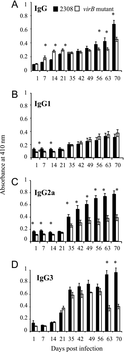
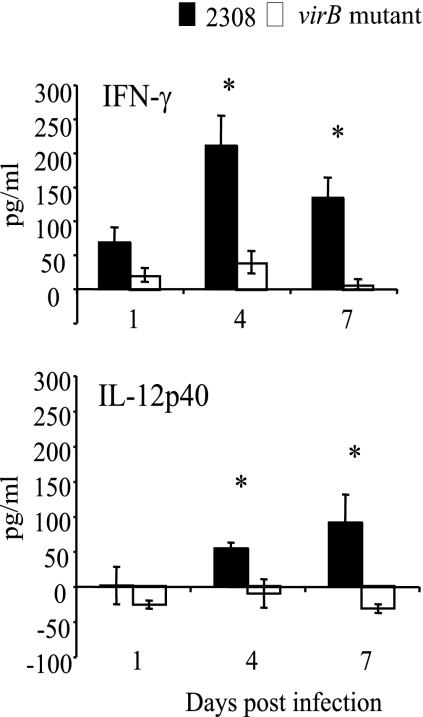
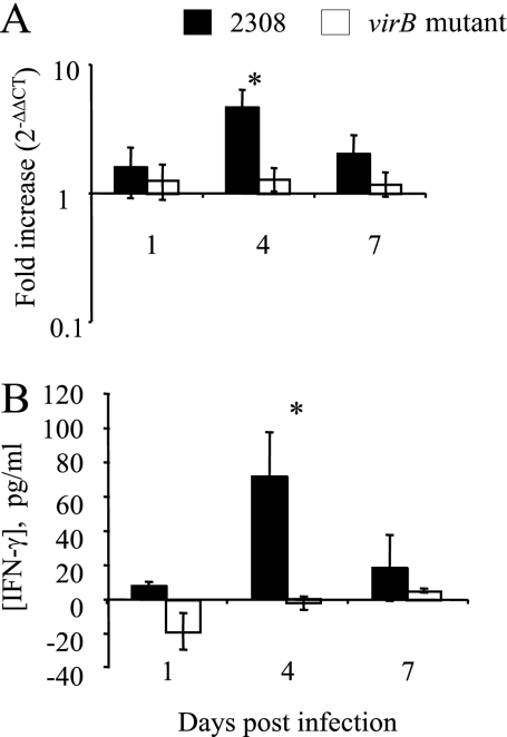
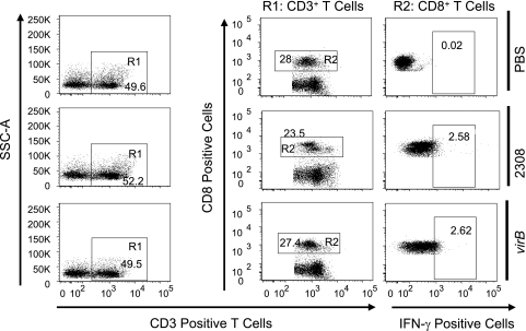
The Brucella abortus type IV secretion system (T4SS), encoded by the virB operon, is essential for establishing persistent infection in the murine reticuloendothelial system. To gain insight into the in vivo interactions mediated by the T4SS, we compared host responses elicited by B. abortus with those of an isogenic mutant in the virB operon. Mice infected with the B. abortus virB mutant elicited smaller increases in serum levels of immunoglobulin G2a, gamma interferon (IFN-gamma), and interleukin-12p40 than did mice infected with wild-type B. abortus. Despite equal bacterial loads in the spleen, at 3 to 4 days postinfection, levels of IFN-gamma were higher in mice infected with wild-type B. abortus than in mice infected with the virB mutant, as shown by real-time PCR, intracellular cytokine staining, and cytokine levels. IFN-gamma-producing CD4(+) T cells were more abundant in spleens of mice infected with wild-type B. abortus than in virB mutant-infected mice. Similar numbers of IFN-gamma-secreting CD8(+) T cells were observed in the spleens of mice infected with B. abortus 2308 or a virB mutant. These results suggest that early differences in cytokine responses contribute to a stronger Th1 polarization of the immune response in mice infected with wild-type B. abortus than in mice infected with the virB mutant.


Figures

FIG. 1.
FIG. 1.
Measurement of antibody levels in sera from mice infected with B. abortus 2308 or the virB mutant by ELISA. Samples from groups of 10 mice inoculated i.p. with 5 × 10⁵ cells of either B. abortus 2308 or the virB mutant were taken at different times postinoculation. The significance of differences between arithmetic means of data for wild-type and mutant mice at each time point was determined using a Student's t test. Asterisks indicate significant differences between immunoglobulin levels in mice infected with 2308 and those in mice infected with the virB mutant (P < 0.05).
FIG. 2.
FIG. 2.
Quantification of cytokine levels in sera of mice infected with B. abortus 2308 or the virB mutant. Samples from groups of 10 mice infected i.p. with either B. abortus 2308 or the virB mutant were taken at different times postinoculation, and cytokine concentrations were determined by Multi-Plex assays. Data shown are increases in cytokine concentrations over preinfection levels. The significance of differences for wild-type and mutant mice at each time point was determined using a paired Student's t test. Error bars represent standard deviations. Asterisks indicate significant differences between the wild type and the mutant (P < 0.05).
FIG. 3.
FIG. 3.
Quantification of IFN-γ levels in spleens of mice infected with B. abortus 2308 or the virB mutant. (A) Transcript levels in spleens from groups of five mice infected with either B. abortus 2308 (black bars) or the virB mutant (white bars) were assayed by real-time reverse transcription-PCR at different times postinoculation. Increases (2−ΔΔCT) in transcript levels were calculated by normalizing CT values for IFN-γ levels to CT values for GAPDH levels and to CT values for IFN-γ levels measured in a group of mock-infected mice. (B) Cytokine measurement of IFN-γ in splenic tissue by Multi-Plex assay was performed with groups of five mice infected with either B. abortus 2308 or the virB mutant at different times postinoculation. Data shown are increases in cytokine concentrations over preinfection levels. The significance of differences for wild-type and mutant mice at each time point was determined using a paired Student's t test. Error bars represent standard deviations. Asterisks indicate significant differences between the wild type and the mutant (P < 0.05). Results shown at the top and bottom are from two independent experiments.
FIG. 4.
FIG. 4.
IFN-γ-producing CD4+ T cells in spleens of mice after infection with B. abortus 2308 or the virB mutant. C57BL/6 mice (four mice per group) were injected i.p. with PBS or 5 × 10⁵ CFU of B. abortus 2308 or the virB mutant. Mice were sacrificed 3 days after infection, and splenocytes were stained with a cocktail of Pacific Blue-conjugated anti-mouse CD4 (L3T4), Alexa Fluor 700-conjugated anti-mouse CD8a (Ly-2), APC-Alexa Fluor 750 anti-mouse CD3e, and phycoerythrin-conjugated anti-mouse IFN-γ. One million cells/sample were acquired with an LSRII apparatus. Numbers indicate the percentages of cells in each gate. Data for one mouse per group are shown as a representative of the group. SSC, side scatter.
FIG. 5.
FIG. 5.
IFN-γ-producing CD8+ T cells in spleens of mice after infection with B. abortus 2308 or the virB mutant. C57BL/6 mice (four mice per group) were injected i.p. with PBS or 5 × 10⁵ CFU of B. abortus 2308 or the virB mutant and sacrificed 3 days after infection. Splenocytes were stained as described in the legend of Fig. 4. One million cells/sample were acquired with an LSRII apparatus. Numbers indicate the percentages of cells in each gate. Data for one mouse per group are shown as a representative of the group. SSC, side scatter.
FIG. 6.
FIG. 6.
Percentage of splenic CD4+ T cells or CD8+ T cells secreting IFN-γ after infection with B. abortus 2308 or the virB mutant. C57BL/6 mice (four mice per group) were injected i.p. with PBS or 5 × 10⁵ CFU of B. abortus 2308 or the virB mutant. Mice were sacrificed 3 days after infection, and cells/mouse were stained for IFN-γ-producing cells. One million cells/sample were acquired with an LSRII apparatus. The asterisk represents statistical significance determined using a Student's t test on data after logarithmic conversion.

Similar articles

Cited by

References

    1. Agranovich, I., D. E. Scott, D. Terle, K. Lee, and B. Golding. 1999. Down-regulation of Th2 responses by Brucella abortus, a strong Th1 stimulus, correlates with alterations in the B7.2-CD28 pathway. Infect. Immun. 674418-4426. - PMC - PubMed
    1. Alton, G. G., L. M. Jones, and D. E. Pietz. 1975. Laboratory techniques in brucellosis. Monogr. Ser. World Health Organ. 1-163. - PubMed
    1. Araj, G. F. 1999. Human brucellosis: a classical infectious disease with persistent diagnostic challenges. Clin. Lab. Sci. 12207-212. - PubMed
    1. Branden, L. J., and C. I Smith. 2002. Bioplex technology: novel synthetic gene delivery system based on peptides anchored to nucleic acids. Methods Enzymol. 346106-124. - PubMed
    1. Caron, E., T. Peyrard, S. Kohler, S. Cabane, J. P. Liautard, and J. Dornand. 1994. Live Brucella spp. fail to induce tumor necrosis factor alpha excretion upon infection of U937-derived phagocytes. Infect. Immun. 625267-5274. - PMC - PubMed

Publication types