. 2008 Jul;76(7):3011-8.
doi: 10.1128/IAI.01672-07. Epub 2008 May 5.

Expression of CD1d and ligand-induced cytokine production are tissue specific in mucosal epithelia of the human lower reproductive tract

Affiliations

Expression of CD1d and ligand-induced cytokine production are tissue specific in mucosal epithelia of the human lower reproductive tract

Kei Kawana et al. Infect Immun. 2008 Jul.

Abstract

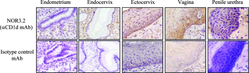
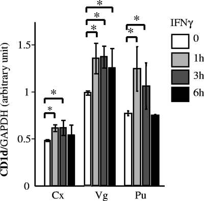
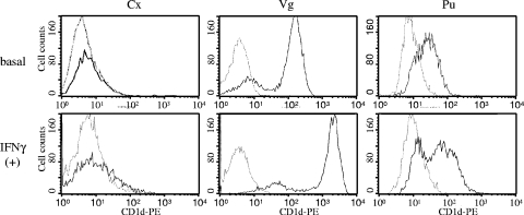
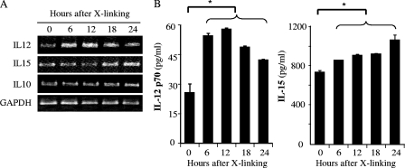
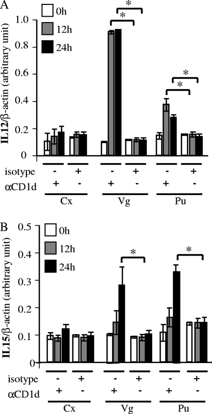
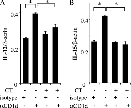
Mucosal epithelia of the human lower reproductive tract (vagina, cervix, and penile urethra) are exposed to sexually transmitted microbes, including Chlamydia trachomatis. The in vivo susceptibility of each tissue type to infection with C. trachomatis is quite distinct. CD1d is expressed on the surface of antigen-presenting cells, including mucosal epithelial cells, and interacts specifically with invariant NKT cells. Invariant NKT cells play a role in both innate and adaptive immune responses to microbes. Here we assessed CD1d expression in normal reproductive tissues by using immunohistochemistry. Immortalized epithelial cell lines from the human lower reproductive tract (vagina, endocervix, and penile urethra) were examined for CD1d expression and for ligand-induced cytokine production induced by CD1d cross-linking. CD1d expression in normal tissue was strong in the vagina but weak in the endocervix and penile urethra. Gamma interferon exposure induced CD1d transcription in all of the cell types studied, with the strongest induction in vaginal cells. Flow cytometry revealed cell surface expression of CD1d in vaginal and penile urethral epithelial cells but not in endocervical cells. Ligation of surface-expressed CD1d by monoclonal antibody cross-linking promoted interleukin-12 (IL-12) and IL-15, but not IL-10, production in vaginal and penile urethral cells. No induction was demonstrated in endocervical cells. CD1d-mediated cytokine production in penile urethral cells was abrogated by C. trachomatis infection. Basal deficiency in CD1d-mediated immune responsiveness may result in susceptibility to sexually transmitted agents. Decreased CD1d-mediated signaling may help C. trachomatis evade detection by innate immune cells.


Figures

FIG. 1.
FIG. 1.
Immunostaining of human normal reproductive tract epithelia for CD1d. Immunostaining for CD1d was performed on formalin-fixed, paraffin-embedded tissue sections of normal human endocervix, ectocervix, vagina, penile urethra, and proliferative-phase endometrium after antigen retrieval. CD1d was detected with NOR3.2, a CD1d-specific MAb (1:500) (upper panels). An isotype-matched control MAb was used as a negative control (lower panels) (magnification, ×200). Results are representative of 2 to 10 normal tissue samples from each site.
FIG. 2.
FIG. 2.
CD1d expression in human reproductive tract epithelial cell lines. Immortalized epithelial cell lines derived from the cervix (Cx), vagina (Vg), and penile urethra (Pu) were cultured with or without IFN-γ (100 ng/ml) for 1 to 6 h. cDNA was produced via RT of 1 μg of total RNA extracted from these cells and then amplified by PCR with primer pairs set in the CD1d and GAPDH genes. Expected single-band PCR products were quantitated with an image analyzer (Scion Image, Frederick, MD) and normalized to GAPDH. Mean values with standard deviations are presented. Asterisks indicate comparisons (exposure versus nonexposure) with statistical significance (P < 0.05; n = 6).
FIG. 3.
FIG. 3.
Cell surface expression of CD1d in human reproductive tract epithelial cell lines. Immortalized epithelial cell lines derived from the cervix (Cx), vagina (Vg), and penile urethra (Pu) were cultured with or without IFN-γ (100 ng/ml) for 24 h. The cells were stained with the anti-CD1d MAb NOR3.2 and a phycoerythrin (PE)-conjugated goat anti-mouse Ig secondary antibody (bold line). Background level staining of the cells with an isotype-matched control antibody is also shown (thin line). The upper and lower panels show cell surface CD1d levels in nontreated (basal) and IFN-γ-treated cells [IFN-γ (+)]. The histograms shown are representative of at least three separate experiments.
FIG. 4.
FIG. 4.
Autocrine cytokines production by CD1d cross-linking in penile urethral epithelial cells. Ten micrograms per milliliter anti-CD1d MAb (51.1) was added to a monolayer of epithelial cells and incubated for 1 h. After washing with PBS, 10 μg/ml goat anti-mouse Ig antibody was added as a cross-linker for 30 min. The cells were incubated in serum-free growth medium without any antibiotics for 0 to 24 h. (A) cDNA was produced via RT of 1 μg of total RNA extracted and amplified by PCR with primer pairs for IL-10, IL-12 p35, IL-15, and GAPDH. PCR products were separated over an ethidium bromide-containing agarose gel. (B) Autocrine cytokine secretion from the epithelial cell at each time point was assessed by ELISA for IL-12 p70 and IL-15. Mean values with standard deviations are presented. Asterisks indicate comparisons (before versus after cross-linking [X-linking]) with statistical significance (P < 0.05; n = 4).
FIG. 5.
FIG. 5.
Quantitative analysis of cytokine production after CD1d cross-linking. CD1d cross-linking was performed as described in the legend to Fig. 4. Anti-CD1d 51.1 (αCD1d) and isotype-matched control (isotype) MAbs were used as primary antibodies for cross-linking. Cells were harvested at 0, 12, and 24 h after cross-linking. IL-12 p35 (A) and IL-15 (B) mRNA levels were analyzed by quantitative RT-PCR by SYBR green methodology. IL-12 p35 and IL-15 mRNA levels were normalized to the β-actin mRNA level. Mean mRNA levels and standard deviations are plotted against time. Asterisks indicate comparisons (isotype versus αCD1d at each time point) with statistical significance (P < 0.05; n = 6). Cx, cervix; Vg, vagina; Pu, penile urethra.
FIG. 6.
FIG. 6.
Abrogation of autocrine cytokine production via CD1d intracellular signaling. Penile urethral epithelial cells were infected (CT+) with C. trachomatis serovar F (multiplicity of infection = 1, 70% infection) and harvested at 24 h postinfection. Control cells (CT−) remained uninfected. Thereafter, antibody cross-linking with the isotype-matched control (isotype) or 51.1 (αCD1d) MAb was performed as described in the legend to Fig. 5. The cells were harvested 12 h after cross-linking. The levels of IL-12 p35 (A) and IL-15 (B) mRNAs were analyzed by quantitative RT-PCR and normalized to β-actin. Mean values with standard deviations are presented. Asterisks indicate comparisons (isotype versus αCD1d) with statistical significance (P < 0.05; n = 4).

Similar articles

Cited by

References

    1. Andreakos, E., R. O. Williams, J. Wales, B. M. Foxwell, and M. Feldmann. 2006. Activation of NF-κB by the intracellular expression of NF-κB-inducing kinase acts as a powerful vaccine adjuvant. Proc. Natl. Acad. Sci. USA 103:314459-14464. - PMC - PubMed
    1. Behar, S. M., and S. A. Porcelli. 2007. CD1-restricted T cells in host defense to infectious diseases. Curr. Top. Microbiol. Immunol. 314215-250. - PubMed
    1. Bilenki, L., S. Wang, J. Yang, Y. Fan, A. G. Joyee, and X. Yang. 2005. NK T cell activation promotes Chlamydia trachomatis infection in vivo. J. Immunol. 1753197-3206. - PubMed
    1. Blumberg, R. S., C. Terhorst, P. Bleicher, F. V. McDermott, C. H. Allan, S. B. Landau, J. S. Trier, and S. P. Balk. 1991. Expression of a nonpolymorphic MHC class1-like molecule, CD1D, by human intestinal epithelial cells. J. Immunol. 1472518-2524. - PubMed
    1. Bonish, B., D. Jullien, Y. Dutronc, B. B. Huang, R. Modlin, F. M. Spada, S. A. Porcelli, and B. J. Nickoloff. 2000. Overexpression of CD1d by keratinocytes in psoriasis and CD1d-dependent IFN-γ production by NK-T cells. J. Immunol. 1654076-4085. - PubMed

Publication types

MeSH terms

LinkOut - more resources