Comparative Study
. 2008 May;179(1):263-75.
doi: 10.1534/genetics.108.088518. Epub 2008 May 5.

Altered dosage and mislocalization of histone H3 and Cse4p lead to chromosome loss in Saccharomyces cerevisiae

Affiliations
Comparative Study

Altered dosage and mislocalization of histone H3 and Cse4p lead to chromosome loss in Saccharomyces cerevisiae

Wei-Chun Au et al. Genetics. 2008 May.

Abstract

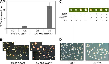
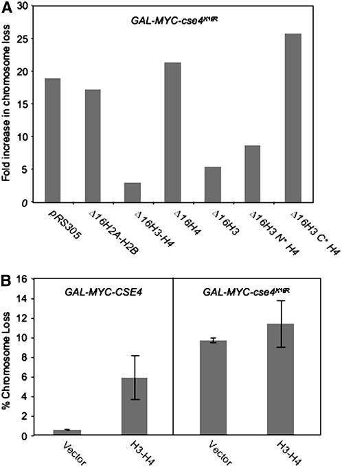
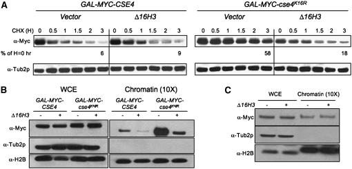
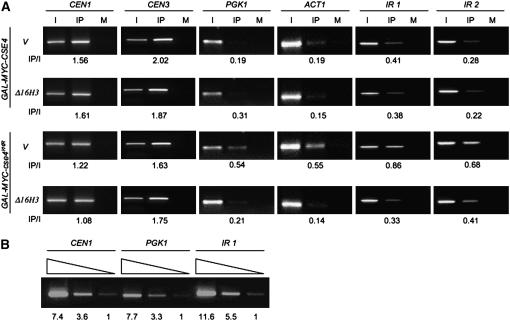
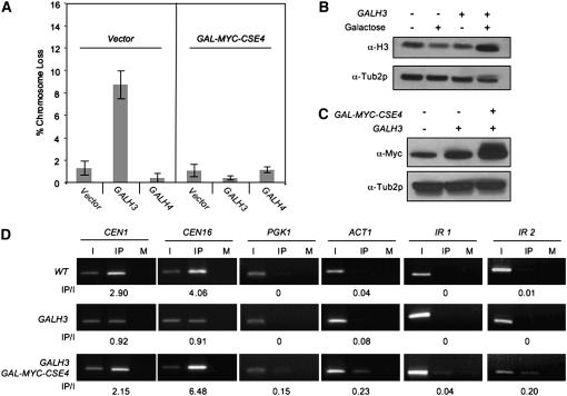
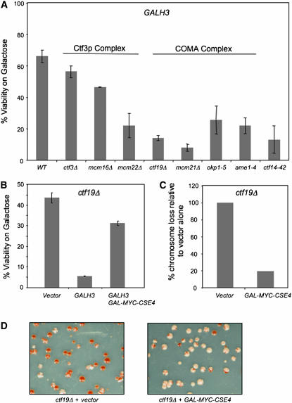
Cse4p is an essential histone H3 variant in Saccharomyces cerevisiae that defines centromere identity and is required for proper segregation of chromosomes. In this study, we investigated phenotypic consequences of Cse4p mislocalization and increased dosage of histone H3 and Cse4p, and established a direct link between histone stoichiometry, mislocalization of Cse4p, and chromosome segregation. Overexpression of the stable Cse4p mutant, cse4(K16R), resulted in its mislocalization, increased association with chromatin, and a high rate of chromosome loss, all of which were suppressed by constitutive expression of histone H3 (delta 16H3). We determined that delta 16H3 did not lead to increased chromosome loss; however, increasing the dosage of histone H3 (GALH3) resulted in significant chromosome loss due to reduced levels of centromere (CEN)-associated Cse4p and synthetic dosage lethality (SDL) in kinetochore mutants. These phenotypes were suppressed by GALCSE4. We conclude that the chromosome missegregation of GALcse4(K16R) and GALH3 strains is due to mislocalization and a functionally compromised kinetochore, respectively. Suppression of these phenotypes by histone delta 16H3 and GALCSE4 supports the conclusion that proper stoichiometry affects the localization of histone H3 and Cse4p and is thus essential for accurate chromosome segregation.


Figures

F<sc>igure</sc> 1.—
Figure 1.—
Expression of GAL-MYC-cse4K16R leads to defects in chromosome transmission fidelity. (A) GAL-MYC-cse4K16R expression leads to a ctf phenotype. YMB3467 and YMB3468 were transformed with GAL-MYC-CSE4 (pSB816) and GAL-MYC-cse4K16R (pSB817) plasmids and plated on SC −URA with limiting adenine plus 2% glucose or 2% galactose/raffinose. The ctf phenotype was quantified by counting the number of colonies that were at least half red for each strain, using three independent transformants. (B) Red sectors in white colonies show the loss of the nonessential chromosome fragment. The inset displays a higher magnification of the colonies of YMB3467 and YMB3468 on 2% galactose/raffinose-containing media. (C) cse4K16R can complement a cse4Δ mutant. cse4K16R∷URA3, in which cse4K16R is expressed from a native CSE4 promoter, was integrated into the cse4Δ/CSE4 heterozygous diploid YMB6267 to make YMB6426, which was sporulated and dissected. The size difference in spores is due to the presence of the reporter chromosome fragment, which causes a slight growth delay. CF, chromosome fragment [CFIII (CEN3L) HIS3 SUP11]. (D) cse4K16R under a native CSE4 promoter does not result in a ctf phenotype. Strains derived from spores (C) containing a wild-type copy of CSE4 or cse4K16R under the endogenous CSE4 promoter (YMB6427) were diluted and plated on SC limiting adenine with 2% glucose.
F<sc>igure</sc> 2.—
Figure 2.—
Constitutive expression of histone H3 from a cell-cycle-independent promoter (Δ16H3) suppresses the GAL-MYC-cse4K16R ctf phenotype. (A) Wild-type strains containing a GAL vector (YMB6108) or expressing GAL-MYC-cse4K16R (YMB6110) were transformed with vector or histone constructs H2A-H2B (pAB95), H3-H4 (pAB157), H4 (pAB203), H3 (pAB204), Δ16H3N*H4 (pAB221), or Δ16H3C*H4 (pAB224) (integrated in the genome at the LEU2 locus), which constitutively express histones from a mutant promoter (Δ16), and were plated on SC limiting adenine with 2% galactose/raffinose. Fold increase in the percentage of colonies that are least half red for GAL-MYC-cse4K16R relative to a wild-type strain with the same integrated histone construct is plotted. The results represent an average of two independent experiments with variation of <5%. (B) The GAL-MYC-cse4K16R ctf phenotype is not suppressed by 2μ H3-H4. A reporter strain expressing 2μ H3-H4 (YMB6138) was transformed with a GAL vector, GAL-MYC-CSE4, or GAL-MYC-cse4K16R and plated on SC −URA with limiting adenine and 2% galactose/raffinose or 2% glucose. The ctf phenotype was quantified as described in Figure 1A. Values represent the average and standard deviation of chromosome loss for three transformants.
F<sc>igure</sc> 3.—
Figure 3.—
Expression of Δ16H3 reduces the stability and chromatin association of cse4K16R. (A) Increased stability of cse4K16R is reduced by expression of Δ16H3. Western blot analysis was performed with protein extracts of GAL-MYC-CSE4 or GAL-MYC-cse4K16R strains with vector (YMB3467 and YMB3477, respectively) or Δ16H3 (YMB3468 and YMB3478, respectively). Protein samples were prepared from cultures grown in SC −URA with 2% galactose/raffinose medium for 6 hr and then shifted to 2% glucose and treated with cycloheximide (CHX) (10 μg/ml) for various times (H, hours). Blots were probed with α-Myc antibody to detect the epitope-tagged Cse4p or cse4K16R and α-Tub2p as a loading control. Blots were quantified by normalizing to Tub2p and setting the level in the untreated sample to 100%. (B) Expression of Δ16H3 reduces the level of chromatin-bound cse4K16R. Western blot analysis of whole-cell extract (WCE) or chromatin fractions of strains used in A were prepared after overnight growth in SC −URA with 2% galactose/raffinose. The blot was probed with α-Myc to detect epitope-tagged Cse4p and cse4K16R, and α-Tub2p and α-H2B as loading controls, for the WCE and chromatin, respectively. The chromatin fraction was prepared from 10-fold more cells than were used to prepare the WCE. (C) Expression of Δ16H3 does not result in altered levels of chromatin-bound Cse4p. Western blot analysis of WCE and chromatin fraction was carried out with wild-type strains expressing MYC-CSE4 from its native promoter with either vector (YMB6147) or integrated Δ16H3 (YMB6146) grown to logarithmic phase in YPD. The blot was probed with α-Myc to detect epitope-tagged Cse4p, and α-Tub2p and α-H2B as loading controls, for the WCE and chromatin, respectively.
F<sc>igure</sc> 4.—
Figure 4.—
cse4K16R mislocalizes to noncentromeric DNA and Δ16H3 suppresses this phenotype. (A) ChIP analysis was done using chromatin extracted from strains expressing GAL-MYC-CSE4 or GAL-MYC-cse4K16R with vector (YMB3467 and YMB3477, respectively) or Δ16H3 (YMB3468 and YMB3478, respectively) grown overnight in SC −URA with 2% galactose/raffinose. PCR analysis was done using input (I) and either α-Myc (IP) or α-GST (mock, M) immunoprecipitated DNA as a template for amplifying CEN1, CEN3, PGK1, ACT1, and two AT-rich intergenic regions IR1 (chromosome V, 2,227,624–2,227,965) and IR2 (chromosome III, 163,364–163,695). Fivefold more DNA was used for IP and M samples. PCR conditions using serially diluted input DNA as template were determined for linear amplification. The values for the IP/I are as shown below each row. (B) Verification of the linear range of PCR amplification for ChIP analysis. Input DNA from YMB3467 was fivefold serially diluted and used for PCR amplification of CEN1, PGK1, and IR1 DNA regions. The relative amount of PCR products normalized to the lowest input (designated as 1.0) is shown.
F<sc>igure</sc> 5.—
Figure 5.—
Phenotypes of overexpressed histone H3 (GALH3) are suppressed by GAL-MYC-CSE4. (A) Overexpression of histone H3 results in a ctf phenotype. Wild-type (YMB3482) or GALCSE4 (YMB3467) strains were transformed with vector (pRS424GAL1), GALH3 (pMB1159), or GALH4 (pMB1158) and plated on SC −URA limiting adenine with 2% galactose/raffinose. The ctf phenotype was quantified by counting the number of colonies that were at least half red from three independent transformants. (B) Histone H3 is expressed at a higher level in a GALH3 strain. Western blot analysis was carried out to detect histone H3 levels in the WCEs of WT cells containing empty GAL vector (YMB6132) or expressing GALH3 (YMB6133) in the presence of 2% glucose or 2% galactose/raffinose from overnight cultures. Tub2p is used as a loading control. (C) Overexpression of histone H3 does not alter Myc-Cse4p levels. Western blot analysis was done using whole-cell extracts with strains containing vector (YMB6132), GALH3 (YMB6133), or GALH3 and GALCSE4 (YMB6136) expressing epitope-tagged Cse4p (MYC-CSE4) from its native promoter in a wild-type strain. The strains were grown to logarithmic phase in SC medium with 2% galactose/raffinose. The blot was probed with α-Myc to detect epitope-tagged Cse4p and α-Tub2p as a loading control. (D) GALH3 leads to reduction in CEN-associated Cse4p. ChIP analysis was carried out using strains described in B. The strains were grown to logarithmic phase in SC with 2% galactose/raffinose. Input (I), immunoprecipitated (IP), and mock (M) samples were used for PCR amplification using primers specific for the loci indicated. PCR conditions identical to Figure 4B were used to obtain linear amplification of targeted chromosomal loci. Values of IP/I are shown below each row.
F<sc>igure</sc> 6.—
Figure 6.—
Expression of GALH3 is lethal in kinetochore mutants. (A) A wild-type strain (Y5563) and kinetochore mutants ctf3Δ (YPH1712), mcm16Δ (YPH1714), mcm22Δ (YPH1716), ctf14-42 (YMB372), ctf19Δ (YPH1713), mcm21Δ (YPH1715), okp1-5 (YPH1678), and ame1-4 (YPH1676) were transformed with 2μ GALH3 (pLG41) and plated on SC −URA with either 2% glucose or 2% galactose/raffinose. Percentage of viability was calculated by the ratio of viable colonies on galactose/raffinose media vs. glucose media for a least three independent transformants. (B) GAL-MYC-CSE4 suppresses the synthetic lethality of GALH3 in ctf19Δ strains. ctf19Δ containing vector (pRS425GAL1), GALH3 (pLG41), or GALH3 GAL-MYC-CSE4 (pMB1147) were plated on SC −TRP −URA with 2% glucose or 2% galactose/raffinose. Percentage of viability was determined as described in A. (C) GAL-MYC-CSE4 suppresses the chromosome loss defect of GALH3 in ctf19Δ strains. ctf19Δ containing vector (pRS315) or GAL-MYC-CSE4 (pMB1147) were plated on SC −LEU with 2% glucose or 2% galactose/raffinose media. Chromosome loss was determined as described in Figure 1A. Values plotted are based on setting the chromosome loss phenotype of the ctf19Δ strain with vector alone to 100%. (D) Representative results of the colony-sectoring assay using strains described in C.

Similar articles

Cited by

References

    1. Baker, R. E., K. Harris and K. Zhang, 1998. Mutations synthetically lethal with cep1 target S. cerevisiae kinetochore components. Genetics 149 73–85. - PMC - PubMed
    1. Basrai, M. A., J. Kingsbury, D. Koshland, F. Spencer and P. Hieter, 1996. Faithful chromosome transmission requires Spt4p, a putative regulator of chromatin structure in Saccharomyces cerevisiae. Mol. Cell. Biol. 16 2838–2847. - PMC - PubMed
    1. Black, B. E., M. A. Brock, S. Bedard, V. L. Woods, Jr. and D. W. Cleveland, 2007. An epigenetic mark generated by the incorporation of CENP-A into centromeric nucleosomes. Proc. Natl. Acad. Sci. USA 104 5008–5013. - PMC - PubMed
    1. Bloom, K. S., and J. Carbon, 1982. Yeast centromere DNA is in a unique and highly ordered structure in chromosomes and small circular minichromosomes. Cell 29 305–317. - PubMed
    1. Blower, M. D., and G. H. Karpen, 2001. The role of Drosophila CID in kinetochore formation, cell-cycle progression and heterochromatin interactions. Nat. Cell Biol. 3 730–739. - PMC - PubMed

Publication types

MeSH terms