. 2008 May 5;181(3):461-73.
doi: 10.1083/jcb.200711145.

Pitx2 is functionally important in the early stages of vascular smooth muscle cell differentiation

Affiliations

Pitx2 is functionally important in the early stages of vascular smooth muscle cell differentiation

Yueting Shang et al. J Cell Biol. .

Abstract

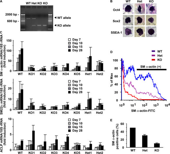
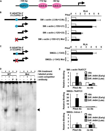
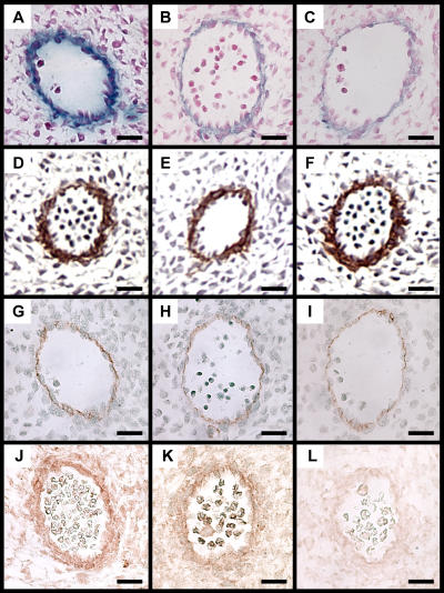
Mechanisms that control vascular smooth muscle cell (SMC) differentiation are poorly understood. We identify Pitx2 as a previously unknown homeodomain transcription factor that is rapidly induced in an in vitro model of SMC differentiation from multipotent stem cells. Pitx2 induces expression of multiple SMC differentiation marker genes by binding to a TAATC(C/T) cis-element, by interacting with serum response factor, and by increasing histone acetylation levels within the promoters of SMC differentiation marker genes. Suppression of Pitx2 reduces expression of SMC differentiation marker genes in the early stages of SMC differentiation in vitro, whereas Prx1, another homeodomain protein, regulates SMC differentiation marker genes in fully differentiated SMCs. Pitx2, but not Prx1, knockout mouse embryos exhibit impaired induction of SMC differentiation markers in the dorsal aorta and branchial arch arteries. Our results demonstrate that Pitx2 functions to regulate the early stages of SMC differentiation.


Figures

Figure 1.
Figure 1.
Pitx2 is induced during SMC differentiation. (A–C) A404 cells were induced to differentiate into SMCs by treatment with 1 μg/ml RA, followed by 0.5-μg/ml puromycin treatment. Expression of Pitx2 (A), SM α-actin (B), and Prx1 (C) was determined by real-time RT-PCR from day (D) 0 to day 7. (D) Expression of Pitx2 isoforms was determined by semiquantitative RT-PCR in undifferentiated A404 cells and RA-treated A404 cells for 48 h. PCR products are 424 bp for Pitx2a, 297 bp for Pitx2b, 234 bp for Pitx2c, and 230 bp for GAPDH. (E and F) ES cells were induced to differentiate into SMCs in an embryoid body system, and expression of Pitx2 (E) and SM α-actin (F) was determined by real-time RT-PCR at days 7, 15, and 28. Values represent the mean ± SEM of three independent experiments.
Figure 2.
Figure 2.
Pitx2 induces expression of SMC differentiation marker genes. (A) Luciferase assays were performed in undifferentiated A404 cells cotransfected with expression plasmids for Pitx2 isoforms and the SM α-actin promoter-enhancer-luciferase construct. RLA, relative luciferase activity. (B) Undifferentiated A404 cells were infected with adenovirus expressing Pitx2a or empty adenovirus, and expression of SMC differentiation marker genes was determined by real-time RT-PCR. (C) Efficiency and specificity of Pitx2 siRNA were examined in COS cells cotransfected with Flag-Pitx2a expression plasmid and Pitx2 siRNA expression plasmid or control plasmids. pMighty-Empty contained no target sequence and pMighty-αScr targeted scrambled sequence. (D) A404 cells were induced to differentiate into SMCs by RA treatment for 1 d (early) or by RA treatment for 3 d followed by puromycin selection for 2 d (late). Cells were transfected with the SM α-actin promoter-enhancer-luciferase construct and a siRNA expression plasmid for Pitx2 (pMighty-αPitx2), Prx1 (pMighty-αPrx1), or pMighty-αScr. Luciferase assays were performed. (E and F) A404 cells were induced to differentiate into SMCs by RA treatment and were transfected with siRNA duplexes for Pitx2 or EGFP. Expression of SMC differentiation marker genes was determined by real-time RT-PCR (E) or Western blotting (F). Values represent the mean ± SEM of three independent experiments.
Figure 3.
Figure 3.
Induction of SMC differentiation marker genes is attenuated in embryoid bodies derived from Pitx2 homozygous knockout ES cells. (A) Genotyping of Pitx2 knockout ES cells. (B) Pitx2 knockout ES cells were stained with Oct4, Sox2, and SSEA-1. (C–E) Five independent Pitx2 homozygous knockout ES cells, two Pitx2 heterozygous knockout ES cells, and wild-type ES cells were induced to differentiate into SMCs in context of embryoid bodies, and expression of SM α-actin, SM22α, and ACLP was determined by real-time RT-PCR (C) and flow cytometry (D and E). Representative data from flow cytometry is shown in D. WT, wild-type; Het, heterozygous; KO, knockout. Values represent the mean ± SEM from three independent experiments.
Figure 4.
Figure 4.
Pitx2 induces SMC differentiation marker genes by binding to the TAATC(C/T) element. (A) Schematic structure of the SM α-actin promoter. CArG B, CArG A, and a TAATCC element are shown. (B and C) Luciferase assays were performed in A404 cells cotransfected with Pitx2a expression plasmid and the promoter-enhancer-luciferase construct of the SM α-actin (B) and SM22α (C) genes. Schematic representation of mutation constructs is shown on the left. Values represent the mean ± SEM from three independent experiments. (D) EMSA was performed using the 32P-labeled double-stranded oligonucleotide containing the TAATCT element of the SM α-actin gene and nuclear extracts from undifferentiated and differentiated A404 cells. Antibodies for Pitx2 (P) or SRF (S) were used to test interference. Arrowhead indicates a labeled oligonucleotide–Pitx2 protein complex. (E) Association of Pitx2 with the TAATC(C/T) region of the SM α-actin and SM22α genes or the first intron region of the SM22α gene was determined by ChIP assays in undifferentiated A404 cells, differentiated A404 cells treated with RA for 2 d (early), and differentiated A404 cells treated with RA for 3 d followed by puromycin selection for 2 d (late). Values represent the mean ± SEM from three independent experiments.
Figure 5.
Figure 5.
Pitx2 and SRF synergistically induce SMC differentiation marker genes. (A) Luciferase assays were performed in A404 cells cotransfected with expression plasmids for Pitx2a and SRF and the SM α-actin promoter-enhancer-luciferase construct. (B) Luciferase assays were performed in A404 cells cotransfected with Pitx2a expression plasmid and mutation constructs of the SM α-actin gene. (C) Coimmunoprecipitation assays were performed in COS cells cotransfected with SRF expression plasmid and wild-type Pitx2a expression plasmid or deletion mutants. Schematic representation of Pitx2a deletion constructs is shown (top). Numbers indicate the position of amino acids. (D) GST pulldown assays were performed using Myc-tagged Pitx2a protein and GST-SRF fusion protein or its deletion mutants. Asterisks indicate GST-SRF protein and its deletion mutants. (E) Mammalian two-hybrid assays were performed in Balb/c 3T3 cells. (F) A404 cells were infected with adenovirus expressing Pitx2a or empty adenovirus, and the association of SRF with the CArG-containing region of the SM α-actin gene was determined by ChIP assays. Values represent the mean ± SEM of three independent experiments. Expression of SRF and GAPDH protein in A404 cells infected with adenovirus expressing Pitx2a or empty adenovirus are also shown.
Figure 6.
Figure 6.
Pitx2 induces acetylation of histone H4 at the promoter regions of SMC differentiation marker genes. (A) A404 cells were infected with adenovirus expressing Pitx2a or empty adenovirus, and acetylation levels of histone H4 at the promoter region of the SM α-actin gene were determined by ChIP assays. (B) Luciferase assays were performed in A404 cells cotransfected with HDAC expression plasmids, Pitx2a expression plasmid, and the SM α-actin promoter-enhancer-luciferase construct. (C, left) Efficiency of p300 siRNA was tested by Western blotting. (C, right) Luciferase assays were performed in A404 cells cotransfected with Pitx2a expression plasmid, expression plasmid for p300 or p300 siRNA, and the SM α-actin promoter-enhancer-luciferase construct. (D) A404 cells were infected with adenovirus expressing Pitx2a or empty adenovirus, and the association of p300, HDAC2, and HDAC5 with the CArG-containing region of the SM α-actin gene was determined by ChIP assays. Values represent the mean ± SEM of three independent experiments.
Figure 7.
Figure 7.
Expression of SMC differentiation markers is nearly abolished in vessels of Pitx2 knockout mouse embryos. (A–O) Expression of SM α-actin (A–C), SM22α (D–F), ACLP (G, I, and K), PECAM (H, J, and L), and Pitx2 (M–O) was examined by immunohistochemistry in wild-type (A, D, G, H, and M), heterozygous Pitx2 knockout (B, E, I, J, and N), and homozygous Pitx2 knockout (C, F, K, L, and O) mouse embryos at E11.5. (P–R) Expression of SM α-actin was examined by immunohistochemistry in wild-type (P), heterozygous Prx1 knockout (Q), and homozygous Prx1 knockout (R) mouse embryos at E11.5. III, third branchial arch artery; IV, fourth branchial arch artery; VI, sixth branchial arch artery; DA, dorsal aorta. Original magnification, 40×. Bars, 100 μm.
Figure 8.
Figure 8.
Expression of SMC differentiation markers is nearly abolished in vessels of Pitx2 knockout mouse embryos. Expression of SM α-actin (A–C), ACLP (D–F), PECAM (G–I), and Pitx2 (J–L) was examined in the dorsal aorta of wild-type (A, D, G, and J), heterozygous Pitx2 knockout (B, E, H, and K), and homozygous Pitx2 knockout (C, F, I, and L) mouse embryos at E11.5. Original magnification, 400×. Bars, 20 μm.

Similar articles

Cited by

References

    1. Ai, D., W. Liu, L. Ma, F. Dong, M.-F. Lu, D. Wang, M.P. Verzi, C. Cai, P.J. Gage, S. Evans, et al. 2006. Pitx2 regulates cardiac left-right asymmetry by patterning second cardiac lineage-derived myocardium. Dev. Biol. 296:437–449. - PMC - PubMed
    1. Bergwerff, M., A.C. Gittenberger-de Groot, L.J. Wisse, M.C. DeRuiter, A. Wessels, J.F. Martin, E.N. Olson, and M.J. Kern. 2000. Loss of function of the Prx1 and Prx2 homeobox genes alters architecture of the great elastic arteries and ductus arteriosus. Virchows Arch. 436:12–19. - PubMed
    1. Carmeliet, P. 2000. Mechanisms of angiogenesis and arteriogenesis. Nat. Med. 6:389–395. - PubMed
    1. Chen, J., C.M. Kitchen, J.W. Streb, and J.M. Miano. 2002. Myocardin: a component of a molecular switch for smooth muscle differentiation. J. Mol. Cell. Cardiol. 34:1345–1356. - PubMed
    1. Drab, M., H. Haller, R. Bychkov, B. Erdmann, C. Lindschau, H. Haase, I. Morano, F.C. Luft, and A.M. Wobus. 1997. From totipotent embryonic stem cells to spontaneously contracting smooth muscle cells: a retinoic acid and db-cAMP in vitro differentiation model. FASEB J. 11:905–915. - PubMed

Publication types

MeSH terms