. 2008 May 5;181(3):475-83.
doi: 10.1083/jcb.200710127.

Cdc14B depletion leads to centriole amplification, and its overexpression prevents unscheduled centriole duplication

Affiliations

Cdc14B depletion leads to centriole amplification, and its overexpression prevents unscheduled centriole duplication

Jun Wu et al. J Cell Biol. .

Abstract

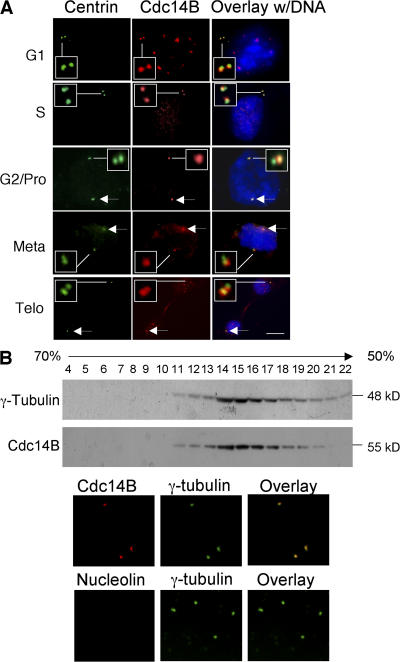
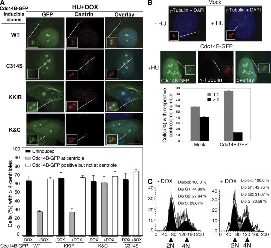
Centrosome duplication is tightly controlled in coordination with DNA replication. The molecular mechanism of centrosome duplication remains unclear. Previous studies found that a fraction of human proline-directed phosphatase Cdc14B associates with centrosomes. However, Cdc14B's involvement in centrosome cycle control has never been explored. Here, we show that depletion of Cdc14B by RNA interference leads to centriole amplification in both HeLa and normal human fibroblast BJ and MRC-5 cells. Induction of Cdc14B expression through a regulatable promoter significantly attenuates centriole amplification in prolonged S phase-arrested cells and proteasome inhibitor Z-L(3)VS-treated cells. This inhibitory function requires centriole-associated Cdc14B catalytic activity. Together, these results suggest a potential function for Cdc14B phosphatase in maintaining the fidelity of centrosome duplication cycle.


Figures

Figure 1.
Figure 1.
A fraction of Cdc14B associates with centrioles. (A) Overlay images depict partial colocalization of endogenous Cdc14B (red) with centrin (green) in U2OS cells during different stages of the cell cycle. Stages of the cell cycle were determined by centrin-labeled centrioles and DAPI-stained DNA (blue). Arrows indicate centrin and Cdc14B-labeled centrioles. Magnified images of centrioles are shown in the insets. Bar, 5 μm. (B, top) Cdc14B cofractionates with γ-tubulin. Lysates of asynchronized HeLa cells were fractionated on a 50–70% sucrose gradient. Fractions 4–22 were analyzed by Western blotting with γ-tubulin and Cdc14B antibodies. (bottom) Cdc14B but not nucleolin (a noncentrosomal protein) costains with γ-tubulin on purified centrosomes (top, fraction 15).
Figure 2.
Figure 2.
Role of phosphatase activity and nucleocytoplasmic transport in Cdc14B centrosome localization. (A) Cdc14B-GFP fusion proteins were induced by DOX for 72 h in U2OS Tet-On stable cell lines carrying different Cdc14B-GFP constructs as indicated. Centrosomes were visualized by γ-tubulin staining (red) and overlaid with Cdc14B-GFP (green) and DAPI-stained DNA (blue). Cdc14BC314S-GFP was not detectable at interphase or mitotic centrosomes (arrows). Insets represent magnified images of centrosomes. Bar, 5 μm. (B) Histogram shows the percentage of interphase and mitotic cells with the indicated Cdc14B-GFP at centrosomes 72 h after DOX addition. The interphase data represent the means ± SD of three independent experiments and at least 500 cells were counted in each experiment. The mitotic experiment was performed in triplicates and a total of 113 Cdc14BC314S-GFP– and 374 Cdc14BWT-GFP–positive cells were counted.
Figure 3.
Figure 3.
Depletion of Cdc14B leads to centriole amplification in HeLa cells. (A) Western blot analysis using an anti-Cdc14B antibody showed Cdc14B knockdown in TREx-HeLa Cdc14BsiRNA stable clones No. 2 and 3 but not in TREx HeLa and clone No. 1. The same Cdc14BsiRNA also led to Cdc14B-GFP knockdown in U2OS Tet-On cells visualized by immunoblotting with anti-GFP antibody. β-actin was used as a loading control. (B) Images of supernumerary centrioles in Cdc14B knockdown HeLa cells. Centrioles were visualized by an anti-centrin antibody. Insets show magnified images of centrioles. Bar, 4 μm. (C) The percentage of cells with more than four centrioles was calculated from the experiments shown in A and B (left). Note that the data shown here does not include polyploid cells described in Fig. S3 A (available at http://www.jcb.org/cgi/content/full/jcb.200710127/DC1). (D, top) Immunoblot shows the level of endogenous Cdc14B and Cdc14A in HeLa cells transfected with siGLO control oligos (lane 1), Cdc14BsiRNA SMARTpool (lane 2), pSuper empty vector (lane 3), and pSuper-Cdc14BshRNA-E9 (lane 4). (bottom) HeLa cells were transfected with the corresponding oligos and vectors as in the top panel. Percentages of cells with more than four centrioles were calculated from experiments shown in B (middle and right). All the data are presented as the means ± SD of three independent experiments. At least 300 cells were counted in each experiment. Note that the indicated fold changes in A and D (top) were calculated based on the densitometric values of each lane normalized against the β-actin loading controls. (E) Supernumerary centrosomes in Hela cells transiently transfected with Cdc14BsiRNA SMARTpool were not the products of centrosome fragmentation. Note that all the γ-tubulin–decorated centrosomes were similar in size and contained centrin-labeled centrioles, and that most of the centrioles were in pairs. Bar, 5 μm.
Figure 4.
Figure 4.
Depletion of Cdc14B causes centriole amplification in normal human fibroblast cells. (A) Representative confocal images of BJ and MRC-5 cells with clustered supernumerary centrioles are shown. Centrioles were labeled by anti-centrin antibody. Insets show magnified images of centrioles. Bar, 10 μm. (B) The percentage of cells with more than four centrioles was calculated from the experiments shown in A. Data shown represent the means ± SD of three independent experiments. At least 300 cells were counted in each experiment.
Figure 5.
Figure 5.
Cdc14B phosphatase activity is required to prevent HU-induced centriole amplification. (A, top) Cdc14B-GFP fusion proteins were induced by 4 μg/ml DOX in the presence of 2 mM HU for 72 h in U2OS Tet-On stable cell lines carrying different Cdc14B-GFP constructs as indicated. Centrioles were visualized by anti-centrin staining (red) and overlaid with Cdc14B-GFP (green) and DAPI (blue). Note that Cdc14BC314S-GFP was not detectable at centrioles (arrow). Insets show magnified images of centrioles. Bar, 5 μm. (bottom) The percentage of cells with more than four centrioles was calculated from both induced (+DOX) and uninduced (−DOX) Cdc14B-GFP stable clones as indicated. Data shown represent the means ± SD of three independent experiments from two individual Cdc14B-GFP stable clones. At least 500 cells were counted in each experiment. (B, top) U2OS Tet-On cells were transfected as indicated. 16 h after transfection, cells were incubated with (+HU) or without (−HU) 2 mM HU and 4 μg/ml DOX for 72 h. Centrosomes were visualized by γ-tubulin staining (red). Representative centrosome amplification was detected in mock-transfected cells after HU treatment but not in pBI-tet-Cdc14BWT-GFP transfected cells where Cdc14BWT-GFP (green) associated with centrosomes. Insets show magnified images of centrosomes. DAPI (blue), DNA. Bar, 5 μm. (bottom) The percentage of cells with the indicated centrosome numbers was calculated from the experiments shown in the top panel. Centrosomes were counted in both mock and Cdc14B-GFP–transfected cells (Cdc14B-GFP–positive at centrosomes). All the data are shown as the means ± SD of three independent experiments. At least 500 cells were counted in each experiment. (C) Representative fluorescence-activated cell sorting profile on cell cycle distribution of DOX-inducible Cdc14B-GFP U2OS Tet-On stable clones. Cells were cultivated in the presence or absence of 4 μg/ml DOX for 72 h. Positions of cells with 2 N and 4 N DNA contents are labeled with arrowheads.
Figure 6.
Figure 6.
Cdc14B phosphatase activity is required to prevent Z-L3-VS–induced centriole overduplication. (A) Dox-inducible U2OS Tet-On cell lines carrying the indicated Cdc14B-GFPs and mock controls were treated with 1 μM Z-L3-VS in the presence or absence of 4 μg/ml Dox for 48 h. Representative centrioles (red) were visualized by an anti-centrin antibody and DNA (blue) was visualized by DAPI. Bar, 5 μM. (B) The percentage of cells with more than four centrioles was calculated from the experiments shown in A and as indicated. Centrioles were counted in mock, uninduced controls (−Dox) and Cdc14B-GFP–induced (+Dox) cells (Cdc14B-GFP–positive at centrioles). All the data are shown as the means ± SD of three independent experiments. At least 400 cells were counted in each experiment.

References

    1. Ashida, S., H. Nakagawa, T. Katagiri, M. Furihata, M. Iiizumi, Y. Anazawa, T. Tsunoda, R. Takata, K. Kasahara, T. Miki, et al. 2004. Molecular features of the transition from prostatic intraepithelial neoplasia (PIN) to prostate cancer: genome-wide gene-expression profiles of prostate cancers and PINs. Cancer Res. 64:5963–5972. - PubMed
    1. Balczon, R., L. Bao, W.E. Zimmer, K. Brown, R.P. Zinkowski, and B.R. Brinkley. 1995. Dissociation of centrosome replication events from cycles of DNA synthesis and mitotic division in hydroxyurea-arrested Chinese hamster ovary cells. J. Cell Biol. 130:105–115. - PMC - PubMed
    1. Bembenek, J., J. Kang, C. Kurischko, B. Li, J.R. Raab, K.D. Belanger, F.C. Luca, and H. Yu. 2005. Crm1-mediated nuclear export of Cdc14 is required for the completion of cytokinesis in budding yeast. Cell Cycle. 4:961–971. - PubMed
    1. Boutros, R., V. Lobjois, and B. Ducommun. 2007. CDC25B involvement in the centrosome duplication cycle and in microtubule nucleation. Cancer Res. 67:11557–11564. - PubMed
    1. Budhu, A.S., and X.W. Wang. 2005. Loading and unloading: orchestrating centrosome duplication and spindle assembly by Ran/Crm1. Cell Cycle. 4:1510–1514. - PMC - PubMed

Publication types

Substances