. 2008 May 13;105(19):7046-51.
doi: 10.1073/pnas.0802463105. Epub 2008 May 5.

Tissue-engineered endothelial and epithelial implants differentially and synergistically regulate airway repair

Affiliations

Tissue-engineered endothelial and epithelial implants differentially and synergistically regulate airway repair

Brett G Zani et al. Proc Natl Acad Sci U S A. .

Abstract

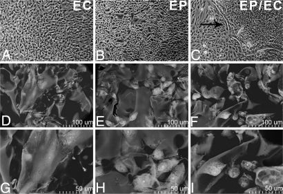
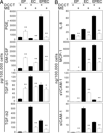
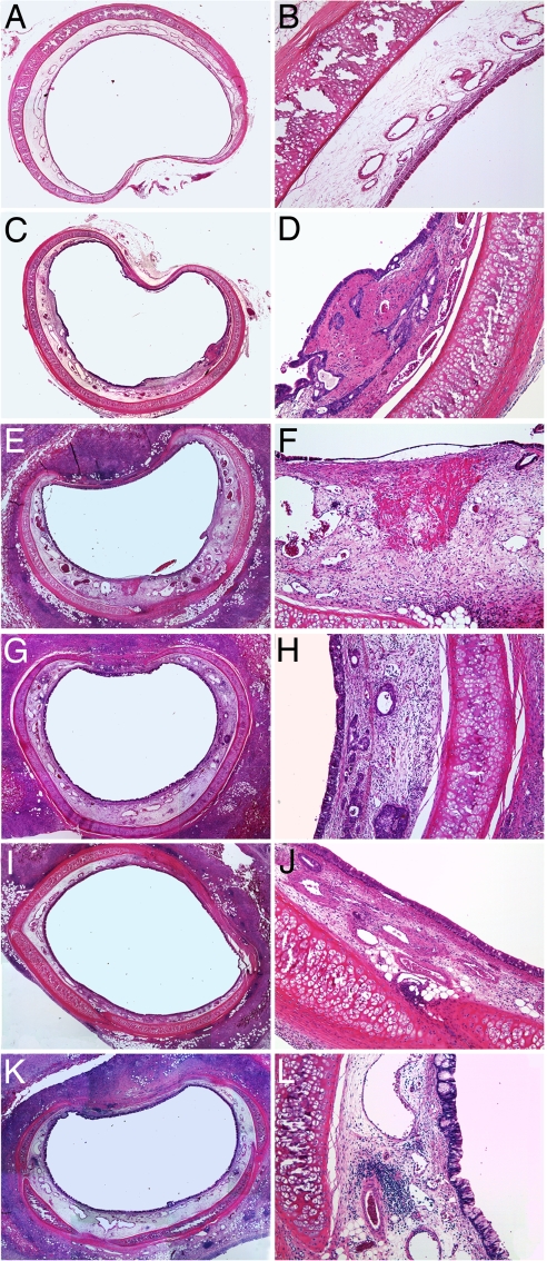
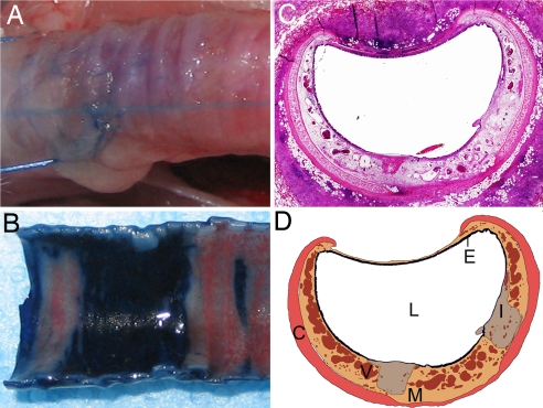
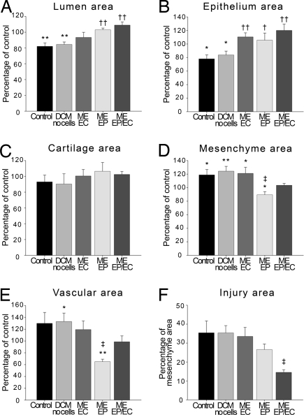
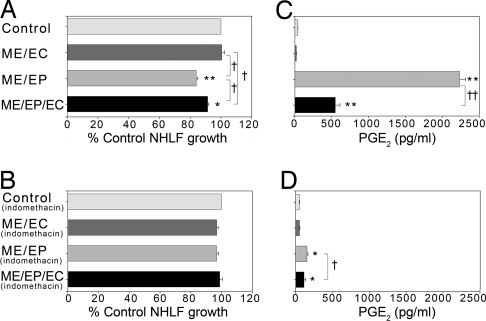
The trilaminate vascular architecture provides biochemical regulation and mechanical integrity. Yet regulatory control can be regained after injury without recapitulating tertiary structure. Tissue-engineered (TE) endothelium controls repair even when placed in the perivascular space of injured vessels. It remains unclear from vascular repair studies whether endothelial implants recapitulate the vascular epithelial lining or expose injured tissues to endothelial cells (ECs) with unique healing potential because ECs line the vascular epithelium and the vasa vasorum. We examined this issue in a nonvascular tubular system, asking whether airway repair is controlled by bronchial epithelial cells (EPs) or by ECs of the perfusing bronchial vasculature. Localized bronchial denuding injury damaged epithelium, narrowed bronchial lumen, and led to mesenchymal cell hyperplasia, hypervascularity, and inflammatory cell infiltration. Peribronchial TE constructs embedded with EPs or ECs limited airway injury, although optimum repair was obtained when both cells were present in TE matrices. EC and EP expression of PGE(2), TGFbeta1, TGFbeta2, GM-CSF, IL-8, MCP-1, and soluble VCAM-1 and ICAM-1 was altered by matrix embedding, but expression was altered most significantly when both cells were present simultaneously. EPs may provide for functional control of organ injury and fibrous response, and ECs may provide for preservation of tissue perfusion and the epithelium in particular. Together the two cells optimize functional restoration and healing, suggesting that multiple cells of a tissue contribute to the differentiated biochemical function and repair of a tissue, but need not assume a fixed, ordered architectural relationship, as in intact tissues, to achieve these effects.


Conflict of interest statement

Conflict of interest statement: The authors have a pending patent on the technology presented in this article that has been licensed to Pervasis Therapeutics. R.L. and E.R.E. hold an equity share in this company.

Figures

Fig. 1.
Fig. 1.
Morphology of EPs, ECs, and EPs/ECs grown on TCPS and with denatured collagen matrices. (A–C) Matrix embedding allows cells to attain a conformation not limited to planar restrictions imposed by TCPS or the apparently distinct isolation of cell types and interposing barrier (black arrow). (Magnification: ×40.) (D–I) Embedded cells conform to the architecture and surface of the local struts of matrix pores in a lesser strained configuration. [Magnification: ×400–450 (D–F) and ×900 (G–I).]
Fig. 2.
Fig. 2.
Protein secretion levels vary depending on cell type and substrate. (A) Protein levels of PGE2, GM-CSF, TGFβ1, and TGFβ2 secreted by EPs, ECs, and EPs/ECs grown on DCCT and matrices were substratum and cell-specific. Cells were grown to confluence and stimulated with 10 ng/ml TNFα for 24 h. (B) Expression of proinflammatory chemokines (IL-8 and MCP-1) and adhesion molecules (sICAM-1 and sVCAM-1) is down-regulated in embedded cells stimulated with 10 ng/ml TNFα for 24 h. *, P < 0.05; **, P < 0.001 vs. DCCT in the same cell type (n = 5–6).
Fig. 3.
Fig. 3.
Peritracheal EP and EC grafts regulate airway injury 9 days after epithelial denuding. (A–F) Injury-induced mesenchymal hyperplasia (C and D) compared with intact controls (A and B) and the acellular implants induced modest peribronchial inflammation (E and F) with more extensive tissue damage, mesenchymal hyperplasia, neovascularization, and luminal narrowing. (G–L) Embedded ECs (G and H) or EPs (I and J) alone reduced these effects and optimally when both cells were present in the constructs (K and L). (Magnification: A, C, E, G, I, and K, ×20; B, D, F, H, J, and L, ×40.)
Fig. 4.
Fig. 4.
Tracheal epithelial injury is localized in the airway injury model. (A and B) Trypan blue is excluded from deposition in the bronchial wall by an intact epithelium (A) but only in areas of remnant epithelium after localized nylon brush injury (B). Areas of epithelial denudation are demarcated by dark blue infiltration. (C and D) Histological cross-sections of rabbit trachea wrapped with a denatured collagen matrix 9 days after injury (C) were digitally reconstructed (D) for quantification of the areas of the lumen (L), epithelium (E), mesenchyme (M), cartilage (C), vascular (V), and injured tissue (I). (Magnification: C, ×20.)
Fig. 5.
Fig. 5.
EP and EC implants promote tracheal healing in a rabbit airway. Although both cell types promote epithelial recovery, EPs protect mesenchymal hyperplasia and prevent neovascularization. Together, the two cells optimize repair. (A–E) Areas measured were lumen (A), epithelium (B), cartilage (C), mesenchyme (D), and vascular (E). (F) Area of tissue injury expressed as percentage of mesenchyme area. Data expressed as mean percentage ± SE of control (noninjured) values from rabbits receiving implants with EPs and ECs (n = 4), only EPs (n = 5), only ECs (n = 7), no cells (n = 7), or no implants (n = 6). *, P < 0.05; **, P < 0.01 vs. control; †, P < 0.05; ††, P < 0.01 vs. acellular matrices and no implants. ‡, P < 0.05 vs. acellular devices, no implants, or embedded ECs.
Fig. 6.
Fig. 6.
PGE2 expression in cocultures of NHLFs and embedded cells inversely correlates with NHLF growth. (A) NHLF growth is reduced in cocultures with EPs embedded alone or with ECs. (B) NHLF growth inhibition is reversed in the same cocultures after 48-h exposure to 5 μM indomethacin. (C) PGE2 levels were higher in NHLF cocultures with EPs embedded alone or with ECs. (D) PGE2 production was significantly diminished after 48-h exposure to 5 μM indomethacin. *, P < 0.00001; **, P < 0.000001 vs. control; †, P < 0.05; ††, P < 0.00001 vs. compared condition (n = 4).

References

    1. Methe H, Edelman ER. Tissue engineering of endothelial cells and the immune response. Transplant Proc. 2006;38:3293–3299. - PMC - PubMed
    1. Methe H, Edelman ER. Cell-matrix contact prevents recognition and damage of endothelial cells in states of heightened immunity. Circulation. 2006;114:I233–I238. - PubMed
    1. Methe H, Groothuis A, Sayegh MH, Edelman ER. Matrix adherence of endothelial cells attenuates immune reactivity: Induction of hyporesponsiveness in allo- and xenogeneic models. FASEB J. 2007;21:1515–1526. - PubMed
    1. Methe H, et al. Matrix embedding alters the immune response against endothelial cells in vitro and in vivo. Circulation. 2005;112:I89–I95. - PubMed
    1. Nathan A, Nugent MA, Edelman ER. Tissue engineered perivascular endothelial cell implants regulate vascular injury. Proc Natl Acad Sci USA. 1995;92:8130–8134. - PMC - PubMed

Publication types

LinkOut - more resources