. 2008 May 13;105(19):6803-8.
doi: 10.1073/pnas.0800019105. Epub 2008 May 5.

Mitochondrial copper(I) transfer from Cox17 to Sco1 is coupled to electron transfer

Affiliations

Mitochondrial copper(I) transfer from Cox17 to Sco1 is coupled to electron transfer

Lucia Banci et al. Proc Natl Acad Sci U S A. .

Abstract

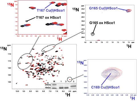
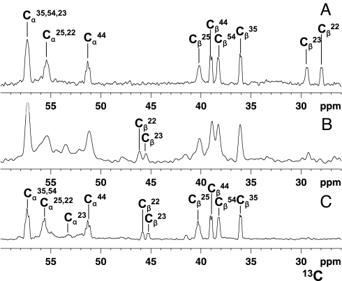
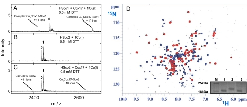
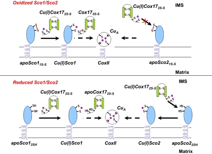
The human protein Cox17 contains three pairs of cysteines. In the mitochondrial intermembrane space (IMS) it exists in a partially oxidized form with two S-S bonds and two reduced cysteines (HCox17(2S-S)). HCox17(2S-S) is involved in copper transfer to the human cochaperones Sco1 and Cox11, which are implicated in the assembly of cytochrome c oxidase. We show here that Cu(I)HCox17(2S-S), i.e., the copper-loaded form of the protein, can transfer simultaneously copper(I) and two electrons to the human cochaperone Sco1 (HSco1) in the oxidized state, i.e., with its metal-binding cysteines forming a disulfide bond. The result is Cu(I)HSco1 and the fully oxidized apoHCox17(3S-S), which can be then reduced by glutathione to apoHCox17(2S-S). The HSco1/HCox17(2S-S) redox reaction is thermodynamically driven by copper transfer. These reactions may occur in vivo because HSco1 can be found in the partially oxidized state within the IMS, consistent with the variable redox properties of the latter compartment. The electron transfer-coupled metallation of HSco1 can be a mechanism within the IMS for an efficient specific transfer of the metal to proteins, where metal-binding thiols are oxidized. The same reaction of copper-electron-coupled transfer does not occur with the human homolog of Sco1, HSco2, for kinetic reasons that may be ascribed to the lack of a specific metal-bridged protein-protein complex, which is instead observed in the Cu(I)HCox17(2S-S)/HSco1 interaction.


Conflict of interest statement

The authors declare no conflict of interest.

Figures

Fig. 1.
Fig. 1.
Titration of 15N-labeled HSco11S-S with unlabeled Cu(I)HCox172S-S followed through chemical shift changes in the 1H–15N HSQC spectra. The 1H–15N HSQC spectrum of 15N-labeled HSco11S-S (in black) is overlaid with the 1H–15N HSQC spectrum of a 1:1 15N-labeled HSco11S-S/unlabeled Cu(I)HCox172S-S mixture (in red). In enlarged views selected regions of 1H–15N HSQC spectra are shown in which also the 1H–15N HSQC spectrum of 15N-labeled Cu(I)HSco1 (in blue) is overlaid. The assignment of the NH resonances of G165 and T167 in Cu(I)HSco1 and HSco11S-S forms is reported. NH resonance of C169 is detected only in the copper(I)-bound form. (Inset) Nonreducing SDS/PAGE gel. Lane M, protein marker; lane 1, HSco1; lane 2, HSco11S-S modified with acetamido-4-maleimidylstilbene-2,2-disulfonic acid (AMS); lane 3, AMS-modified HSco12SH.
Fig. 2.
Fig. 2.
Oxidation state of HCox17 cysteines monitored in the redox reaction between (13C,2H,15N)Cys-selectively labeled Cu(I)HCox172S-S and 15N-labeled HSco11S-S. Chemical shift changes of Cα and Cβ carbons of Cys residues of Cu(I)HCox172S-S were followed by 1D 13C NMR spectra. (A) (13C,2H,15N)Cys-selectively labeled Cu(I)HCox172S-S. (B) 1:1 (13C,2H,15N)Cys-selectively labeled Cu(I)HCox172S-S/15N-labeled HSco11S-S mixture. (C) (13C,2H,15N)Cys-selectively labeled HCox173S-S obtained by air oxidation. The assignment of the Cα and Cβ resonances of the Cys residues of both Cu(I)HCox172S-S and HCox173S-S forms is reported. Cβ signals of the two Cys residues (C22 and C23) of Cu(I)HCox172S-S that are involved in the disulfide exchange reaction drastically reduce their intensity in the protein mixture (B) with the concomitant formation of the corresponding Cβ signals with chemical shifts typical of HCox173S-S.
Fig. 3.
Fig. 3.
Electrospray ionization (ESI) MS analysis of Cu(I)HCox172S-S/HSco protein–protein complex formation in the presence of 0.5 mM DTT (Left) and NMR data on Cu(I)HCox172S-S/HSco21S-S mixture in the absence of reducing agents (Right). (A) Mixture of HSco1 (3 μM), HCox172S-S (2 μM), and Cu(I)DTT (2 μM). (B) Mixture of HSco2 (3 μM) and Cu(I)DTT (2 μM). (C) Mixture of HSco2 (3 μM), HCox172S-S (2 μM), and Cu(I)DTT (2 μM). Conditions: 20 mM ammonium acetate, pH 7.5, 0.5 mM DTT. ESI MS spectra were recorded as described in Materials and Methods. Charge state +8 ions are presented for HSco1 and HSco2, and numbers on the peaks denote the metal stoichiometry of the complex. Peaks corresponding to +10 and +11 ions of HCox172S-S/Cu(I)1/HSco1 complexes (A) and positions of theoretical peaks for HCox172S-S/Cu(I)1/HSco2 complexes (C) are indicated with arrows. (D) 1H–15N HSQC spectrum of a 1:1 15N-labeled HSco22S-S/15N-labeled Cu(I)HCox172S-S mixture (in black) is overlaid with 1H–15N HSQC spectra of 15N-labeled HSco21S-S (in blue) and of 15N-labeled Cu(I)HCox172S-S (in red). The NH resonances of the mixture and of the starting materials are well superimposed and no new NH resonances of Cu(I)HSco1 and apoHCox172S-S species are detected, indicating that no reaction occurs. (Inset) Nonreducing SDS/PAGE gel. Lane M, protein marker; lane 1, HSco2; lane 2, AMS-modified HSco22SH; lane 3, AMS-modified HSco21S-S.
Fig. 4.
Fig. 4.
Proposed mechanism of copper transfer from HCox172S-S to the subunit II of CcO, through the assistance of HSco1 and HSco2. This model implies that HSco1, independently of the redox state of its Cys ligands in the IMS, may accept copper(I) from the mitochondrial chaperone Cu(I)HCox172S-S to form Cu(I)HSco1 whereas HSco2 can accept copper(I) from Cu(I)HCox172S-S only once its Cys ligands in the IMS are in a reduced state. HCox173S-S, produced by the redox HSco1/HCox172S-S reaction, can be quickly reduced to HCox172S-S by GSH, thus the latter protein being recycled for the following metal transfer.

References

    1. Ramirez BE, Malmström BG, Winkler JR, Gray HB. The currents of life: The terminal electron-transfer complex of respiration. Proc Natl Acad Sci USA. 1995;92:11949–11951. - PMC - PubMed
    1. Finney LA, O'Halloran TV. Transition metal speciation in the cell: Insights from the chemistry of metal ion receptors. Science. 2003;300:931–936. - PubMed
    1. Tsukihara T, et al. Structures of metal sites of oxidized bovine heart cytochrome c oxidase at 2.8 Å. Science. 1995;269:1069–1074. - PubMed
    1. Tsukihara T, et al. The whole structure of the 13-subunit oxidized cytochrome c oxidase at 2.8 Å. Science. 1996;272:1136–1144. - PubMed
    1. Carr HS, Winge DR. Assembly of cytochrome c oxidase within the mitochondrion. Acc Chem Res. 2003;36:309–316. - PubMed

Publication types

LinkOut - more resources