. 2008 May 13;105(19):6936-41.
doi: 10.1073/pnas.0710831105. Epub 2008 May 5.

Inhibition of Wnt signaling by the osteoblast-specific transcription factor Osterix

Affiliations

Inhibition of Wnt signaling by the osteoblast-specific transcription factor Osterix

Chi Zhang et al. Proc Natl Acad Sci U S A. .

Abstract

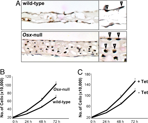
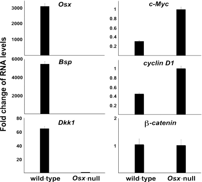
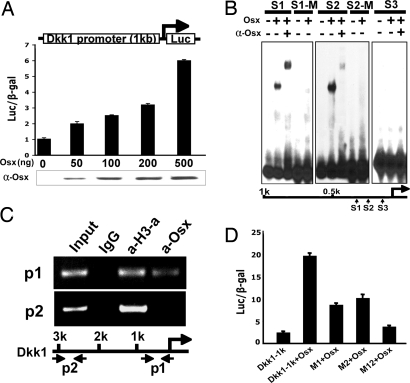
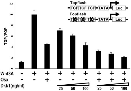
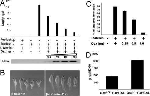
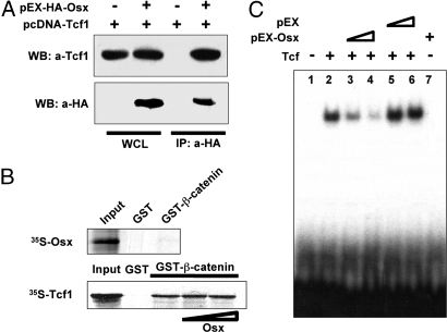
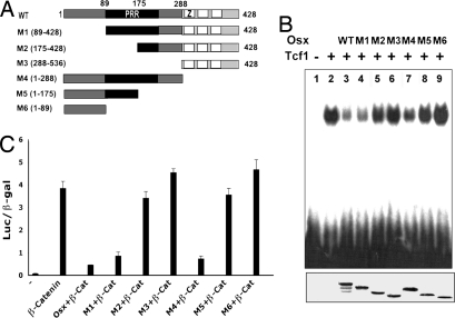
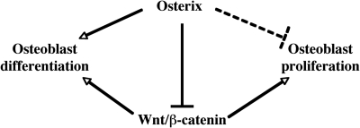
The recent identification of the genes responsible for several human genetic diseases affecting bone homeostasis and the characterization of mouse models for these diseases indicated that canonical Wnt signaling plays a critical role in the control of bone mass. Here, we report that the osteoblast-specific transcription factor Osterix (Osx), which is required for osteoblast differentiation, inhibits Wnt pathway activity. First, in calvarial cells of embryonic day (E)18.5 Osx-null embryos, expression of the Wnt antagonist Dkk1 was abolished, and that of Wnt target genes c-Myc and cyclin D1 was increased. Moreover, our studies demonstrated that Osx bound to and activated the Dkk1 promoter. In addition, Osx inhibited beta-catenin-induced Topflash reporter activity and beta-catenin-induced secondary axis formation in Xenopus embryos. Importantly, in calvaria of E18.5 Osx-null embryos harboring the TOPGAL reporter transgene, beta-galactosidase activity was increased, suggesting that Osx inhibited the Wnt pathway in osteoblasts in vivo. Our data further showed that Osx disrupted binding of Tcf to DNA, providing a likely mechanism for the inhibition by Osx of beta-catenin transcriptional activity. We also showed that Osx decreased osteoblast proliferation. Indeed, E18.5 Osx-null calvaria showed greater BrdU incorporation than wild-type calvaria and that Osx overexpression in C2C12 mesenchymal cells inhibited cell growth. Because Wnt signaling has a major role in stimulating osteoblast proliferation, we speculate that Osx-mediated inhibition of osteoblast proliferation is a consequence of the Osx-mediated control of Wnt/beta-catenin activity. Our results add a layer of control to Wnt/beta-catenin signaling in bone.


Conflict of interest statement

The authors declare no conflict of interest.

Figures

Fig. 1.
Fig. 1.
Effect of Osx on cell proliferation. (A) BrdU incorporation in sections of calvaria from E18.5 wild-type and Osx-null embryos. Pregnant females were injected i.p. with BrdU 4 h before being killed. Sections were obtained from four wild-type and four Osx-null embryos, and BrdU-positive cells were counted in 10 different fields. The percentages of BrdU positive cells were 9.2 ± 1.4 for wild-type calvaria and 45.8 ± 2.8 for Osx-null calvaria. Arrowhead in the insert indicate BrdU-positive cells. (B) Cell growth of E18.5 wild-type and Osx-null primary calvaria cells. (C) A stably transfected C2C12 cell line was generated in which the expression of Osx could be induced by using the Tet-off system in the absence of tetracycline. Comparison of cell growth in the presence (+) or absence (−) of tetracycline.
Fig. 2.
Fig. 2.
Fold change in RNA levels of specific calvaria RNAs from E18.5 Osx wild-type and Osx-null embryos. RNA levels were measured by real-time RT-PCR. The level of each type of RNA from Osx-null calvaria was normalized to a value of 1.
Fig. 3.
Fig. 3.
Osx activates a Dkk1 promoter. (A) HEK293 cells were transfected with a 1-kb Dkk1 promoter luciferase reporter without or with an increasing amount of pEX-Osx as indicated. Cell extracts were subjected to Western blot analysis to monitor the expression of transfected pEX-Osx. (B) Purified recombinant Osx binds to two specific sites in the Dkk1 promoter in EMSA. Three potential Osx binding sites in the 1-kb Dkk1 promoter, S1, S2, and S3, were used as probes. S1-M is S1 site mutation and S2-M is S2 site mutation. (C) Osx associates with the chromatin of a segment of the Dkk1 promoter in ChIP assay. Calvarial cells were isolated and cultured from wild-type new born mice. p1 primer set corresponds to a segment of the Dkk1 promoter covering S1, S2, and S3 sites within the proximal 500 bp. As a negative control, p2 primer set covers a distal region of the Dkk1 promoter, which does not contain an Osx binding site. Anti-acetylated histone H3 antibody (a-H3-a) was used as a positive control. (D) Mutations in sites S1 and S2 inhibit activation of the Dkk1 promoter by Osx. Mutations in the 1-kb Dkk1 reporter were constructed. Mutant M1 contains the S1 site mutation, mutant M2 the S2 site mutation, and double mutant M12 (both S1 and S2 mutations) in the 1-kb Dkk1 promoter. HEK293 cells were transfected with wild-type and mutant Dkk1 promoter reporters without or with pEX-Osx, as indicated.
Fig. 4.
Fig. 4.
Cooperation between Osx and Dkk1 in the inhibition of Wnt3A-induced Topflash activity. Topflash and Fopflash reporters were transfected in HEK293 cells with pEX-Osx as indicated. Wnt3A-conditioned medium was used to activate the Topflash reporter. Recombinant Dkk1 protein was added to the medium at the different concentrations as indicated. Results were expressed as the ratio of Topflash over Fopflash activity.
Fig. 5.
Fig. 5.
Inhibition of β-catenin transcription activity by Osx. (A) Osx inhibits β-catenin-induced Wnt reporter activity. HEK293 cells were transfected with the Topflash or Fopflash reporter along with Myc-β-catenin, a plasmid expressing a stabilized β-catenin, and increasing amounts of pEX-Osx DNA. (B) Osx inhibits β-catenin-induced secondary axis formation in Xenopus embryos. Embryos were microinjected with RNA for stabilized β-catenin (100 pg) and increasing amounts of Osx RNA at the four-cell cleavage stage into the equatorial region of a single vegetal-ventral blastomere. Microinjection of β-catenin RNA in the ventral side of four-cell Xenopus embryos induces the formation of a secondary body axis. The phenotypes were evaluated by using a binocular dissecting microscope at the tadpole stage. (C) Dose-dependent inhibition of β-catenin-induced secondary axis formation in Xenopus embryos by Osx. (D) β-galactosidase activity in calvarial extracts of E18.5 Osx+/+;TOPGAL and Osx−/−;TOPGAL embryos. Results were expressed as the ratio of β- galactosidase over DNA.
Fig. 6.
Fig. 6.
Interaction between Osx and Tcf1. (A) Coimmunoprecipitation (CoIP) of Osx and Tcf1 in transfected HEK293 cells. Here, 1 μg of pEX-HA-Osx and pCDNA-Tcf1 were cotransfected or transfected alone into HEK293 cells. Whole-cell lysates (WCL) were immunoprecipitated with anti-HA (10 μl) and the precipitate was immunoblotted with anti-Tcf1. (B) No disruption of β-catenin interaction with Tcf1 by Osx. (Upper) GST-β-catenin was used to pull down 35S labeled Osx. Ten percent of the synthesized Osx was used as an input, and GST was used as a control. (Lower) GST-β-catenin was used to pull down 35S-labeled Tcf1. Baculovirus-expressed Osx was used as Osx protein source. (C) Disruption of Tcf1 binding to DNA by Osx. Osx and Tcf1 proteins were synthesized by TNT IVTT systems. Tcf1 bound to Tcf 1 binding probe in EMSA. TNT lysates containing pEX-Osx and control TNT lysates with pEX were added to the DNA binding reaction.
Fig. 7.
Fig. 7.
PRR region of Osx is needed for disruption of Tcf1 binding to DNA and for inhibition of β-catenin transcriptional activity. (A) Schematic representation of the Osx deletion mutants. PRR, proline-rich region; Z, zinc-finger domain. (B) Osx and Tcf1 proteins were synthesized by TNT IVTT systems. M1 and M4 are mutants with full PRR region. M2, M3, M5, and M6 are mutants without full PRR region. (Lower) [35S]methionine-labeled Osx wild-type and mutants synthesized by IVTT system were detected by autoradiograph. (C) Osx PRR domain is required for Osx inhibitory effect on Wnt pathway. Vectors expressing Osx wild-type and mutants were transfected in HEK293 cells with or without vectors expressing activated β-catenin (β-cat) with the Topflash reporter.
Fig. 8.
Fig. 8.
Proposed model of coordinated regulation of osteoblast differentiation and proliferation by Osx and Wnt/β-catenin signaling. Wnt/β-catenin signaling has an essential role in osteoblast differentiation during embryonic development and has a major role in stimulating osteoblast proliferation both during embryonic development and postnatally. Osx is an osteoblast-specific transcription factor, required for osteoblast differentiation. The inhibition of Wnt/β-catenin signaling activity by Osx also constitutes a possible mechanism for the inhibition by Osx of osteoblast proliferation. Not shown is the inhibitory role of Wnt/β-catenin in osteoclast differentiation and function.

References

    1. Akiyama H, et al. Osteo-chondroprogenitor cells are derived from Sox9 expressing precursors. Proc Natl Acad Sci USA. 2005;102:14665–14670. - PMC - PubMed
    1. St-Jacques B, Hammerschmidt M, McMahon AP. Indian hedgehog signaling regulates proliferation and differentiation of chondrocytes and is essential for bone formation. Genes Dev. 1999;13:2072–2086. - PMC - PubMed
    1. Komori T, et al. Targeted disruption of Cbfa1 results in a complete lack of bone formation owing to maturational arrest of osteoblasts. Cell. 1997;89:755–764. - PubMed
    1. Mundlos S, et al. Mutations involving the transcription factor CBFA1 cause cleidocranial dysplasia. Cell. 1997;89:773–779. - PubMed
    1. Nakashima K, et al. The novel zinc finger-containing transcription factor osterix is required for osteoblast differentiation and bone formation. Cell. 2002;108:17–29. - PubMed

Publication types

MeSH terms