. 2008 Aug;7(8):1212-25.
doi: 10.4161/cbt.7.8.6220. Epub 2008 Aug 30.

Human breast cancer-associated fibroblasts (CAFs) show caveolin-1 downregulation and RB tumor suppressor functional inactivation: Implications for the response to hormonal therapy

Affiliations

Human breast cancer-associated fibroblasts (CAFs) show caveolin-1 downregulation and RB tumor suppressor functional inactivation: Implications for the response to hormonal therapy

Isabelle Mercier et al. Cancer Biol Ther. 2008 Aug.

Abstract

It is becoming increasingly apparent that the tumor microenvironment plays a critical role in human breast cancer onset and progression. Therefore, we isolated cancer-associated fibroblasts (CAFs) from human breast cancer lesions and studied their properties, as compared with normal mammary fibroblasts (NFs) isolated from the same patient. Here, we demonstrate that 8 out of 11 CAFs show dramatic downregulation of caveolin-1 (Cav-1) protein expression; Cav-1 is a well-established marker that is normally decreased during the oncogenic transformation of fibroblasts. Next, we performed gene expression profiling studies (DNA microarray) and established a CAF gene expression signature. Interestingly, the expression signature associated with CAFs encompasses a large number of genes that are regulated via the RB-pathway. The CAF gene signature is also predictive of poor clinical outcome in breast cancer patients that were treated with tamoxifen mono-therapy, indicating that CAFs may be useful for predicting the response to hormonal therapy. Finally, we show that replacement of Cav-1 expression in CAFs (using a cell-permeable peptide approach) is sufficient to revert their hyper-proliferative phenotype and prevent RB hyper-phosphorylation. Taken together, these studies highlight the critical role of Cav-1 downregulation in maintaining the abnormal phenotype of human breast cancer-associated fibroblasts.


Figures

Figure 1.
Figure 1.
Morphology of breast tissue and mammary stromal fibroblasts. (A) Representative images of H&E stained sections from the tissues used to generate the primary cultures of breast stromal fibroblasts are shown. Upper and lower panels correspond to normal breast tissue and invasive ductal carcinoma (IDC), respectively. The stroma surrounding the invasive ductal carcinoma is highly reactive, containing many fibroblasts, as demonstrated by small and numerous hematoxylin-stained nuclei. All tumors were matched with normal tissue from the same patient. Images were taken with a 20x objective using an Olympus BX51 microscope, a Qimaging Micropublisher 5.0 camera and iVision software. (B) Phase images of primary cultures of fibroblasts isolated from invasive ductal carcinomas (lower) and matching fibroblasts from adjacent normal breast tissue (upper) of the same patient. Fibroblasts isolated from the breast tumor appear more elongated and spindle-shaped. Images were taken at 10x.
Figure 2.
Figure 2.
Downregulation of Cav-1 protein levels in CAFs versus NFs. (A) Fold changes in Cav-1 protein expression in CAFs vs. NFs generated from 11 breast cancer patients. Patient results were divided into three groups: (A), loss of Cav-1 expression; (B), no change in Cav-1 expression; and (C), increased Cav-1 expression. Note that most of the tumor-associated fibroblasts (from 8 of 11 patients) had a reduction in Cav-1 expression (n = 8), while one had no change, and two showed an increase. Thus, we focused our efforts on the patients showing a loss of Cav-1 expression. G corresponds to group and N to number of patients. (B) Immunoblot analysis of Cav-1 expression shows a decrease in Cav-1 levels in cancer-associated fibroblasts (C) when compared to adjacent normal fibroblasts (N). All the tumors analyzed had a matched normal tissue control from the same patient. β-actin was used as a loading control to assure equal loading. 30 μg of total protein lysate was loaded in each lane.
Figure 3.
Figure 3.
CAFs exhibit a hyper-proliferative phenotype. Equal numbers of normal and cancer fibroblasts were plated for 72 hrs and given a two hour pulse of BrdU. Using an ELISA kit, the absorbance was then measured at 370 nm with a reference of 490 nm. The absorbance is reflective of the amount of BrdU incorporated by the cells. CAFs show a ~3.6 fold increase in BrdU incorporation. *p < 0.05. Quantitatively similar results were obtained when BrdU incorporation was measured in CAFs and NFs isolated from several different patients.
Figure 4.
Figure 4.
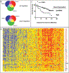
High expression of the breast CAF gene signature is associated with poor clinical outcome in breast cancer patients treated with tamoxifen monotherapy. (A) Venn diagrams summarizing how the two gene signatures were derived by comparing and intersecting the gene sets from matched NFs and CAFs from three different patients. (B) Gene expression data from 60 ER-positive human breast tumors that were both micro- and macrodissected were analyzed for the expression pattern of 118 genes upregulated in CAFs. A core of proliferation associated genes that are regulated by the RB/E2F pathway (marked in red) strongly co-segregated in this analyses. (C) A Kaplan-Meyer survival analysis was conducted, wherein the recurrence of those tumors in the highest quartile of overall expression was compared against the remainder of the cohort (p < 0.001). Patients in the High CAF gene expression group had a poor prognosis on Tamoxifen mono-therapy, with greater than a 3.8-fold reduction in recurrence-free survival.
Figure 5.
Figure 5.
RB phosphorylation and RB-regulated gene products are increased in CAFs. (A) CAFs have increased phosphorylated RB as compared with normal adjacent fibroblasts, as shown by immunufluorescence (Upper panels), using a phopho-specific antibody that recognizes endogeneous RB only when phosphorylated at serine 807/811. (B) CAFs show an increase in the levels of PCNA when compared with normal adjacent fibroblasts, as shown by confocal microscopy (Upper). (C) CAFs show increases in MCM7 expression, as seen by confocal microscopy (Upper). In (A–C), DAPI staining shows the nuclei of the cells imaged (Lower). Images were taken at 20x. Virtually identical results were obtained using CAFs and NFs isolated from several different patients. Representative images are shown.
Figure 6.
Figure 6.
BrdU incorporation is inhibited by a Cav-1 mimetic peptide in CAFs. Equal numbers of cancer-associated fibroblasts were plated for 24 hrs and given a 20 μM dose of a cell-permeable Cav-1 mimetic peptide attached to penetratin. Following 48 hrs of treatment, the cells were given a two hour pulse of BrdU. Using an ELISA kit, the absorbance was then measured at 370 nm with a reference of 490 nm. CAFs show a 3-fold decrease in BrdU incorporation following Cav-1 treatment. *p < 0.05. Equivalent results were obtained using CAFs isolated from several different patients.
Figure 7.
Figure 7.
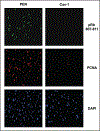
RB-phosphorylation is inhibited by a Cav-1 mimetic peptide in CAFs. Treatment of CAFs with a Cav-1 mimetic peptide inhibited the phosphorylation of RB as shown by immunufluorescence (Upper), using a phospho-specific antibody that recognizes endogeneous RB only when phosphorylated at serine 807/811. PCNA levels were also decreased by the Cav-1 peptide (Middle). Penetratin (Pen) alone did not affect the levels of phospho-RB or PCNA. DAPI staining shows the nuclei of the cells imaged (Lower). Images were taken at 20x. Virtually identical results were obtained with 5, 10 and 20 μM dosages of the Cav-1 mimetic peptide.

Similar articles

Cited by

References

    1. Sappino AP, Skalli O, Jackson B, Schurch W, Gabbiani G. Smooth-muscle differentiation in stromal cells of malignant and non-malignant breast tissues. Int J Cancer 1988; 41:707–12. - PubMed
    1. Ronnov-Jessen L, Petersen OW, Bissell MJ. Cellular changes involved in conversion of normal to malignant breast: importance of the stromal reaction. Physiol Rev 1996; 76:69–125. - PubMed
    1. Tlsty TD, Hein PW. Know thy neighbor: stromal cells can contribute oncogenic signals. Curr Opin Genet Dev 2001; 11:54–9. - PubMed
    1. Mueller MM, Fusenig NE. Friends or foes—bipolar effects of the tumour stroma in cancer. Nat Rev Cancer 2004; 4:839–49. - PubMed
    1. Serini G, Gabbiani G. Mechanisms of myofibroblast activity and phenotypic modulation. Exp Cell Res 1999; 250:273–83. - PubMed

Publication types