. 2006 May;2(2):93-103.
doi: 10.1017/S1740925X0600010X.

Electric field-induced astrocyte alignment directs neurite outgrowth

Affiliations

Electric field-induced astrocyte alignment directs neurite outgrowth

John K Alexander et al. Neuron Glia Biol. 2006 May.

Abstract

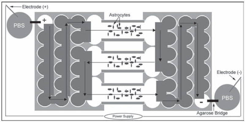
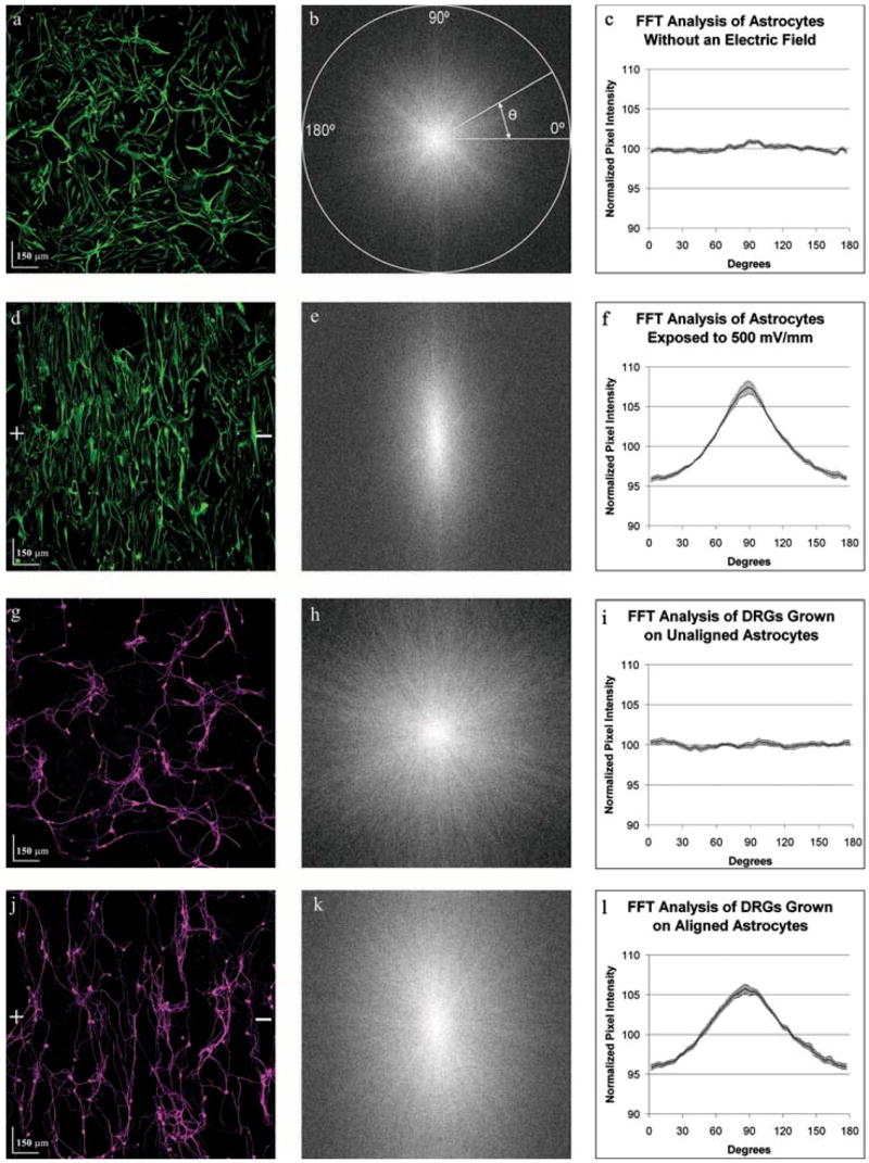
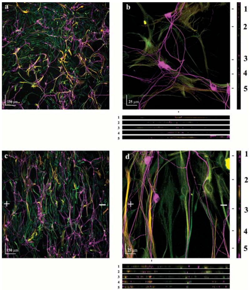
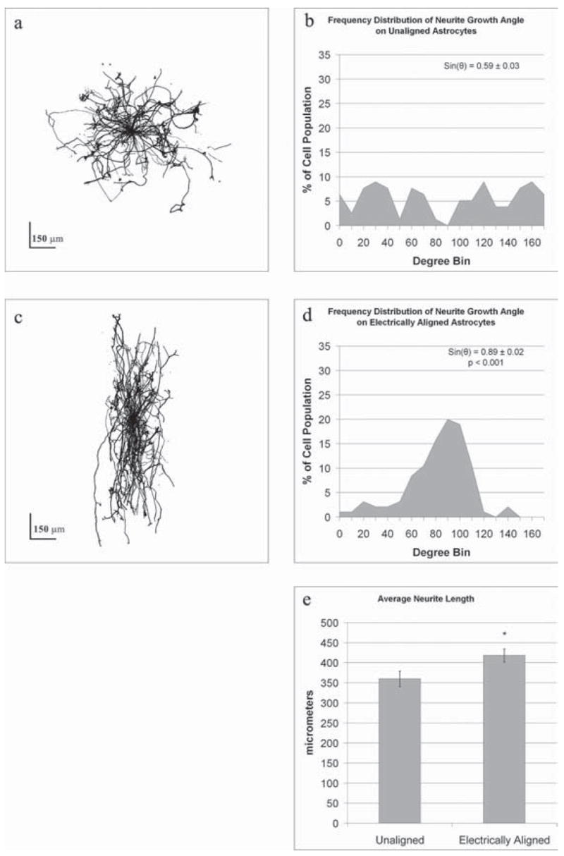
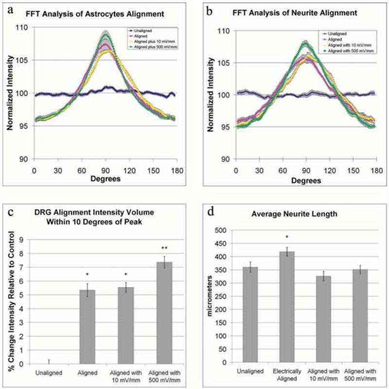
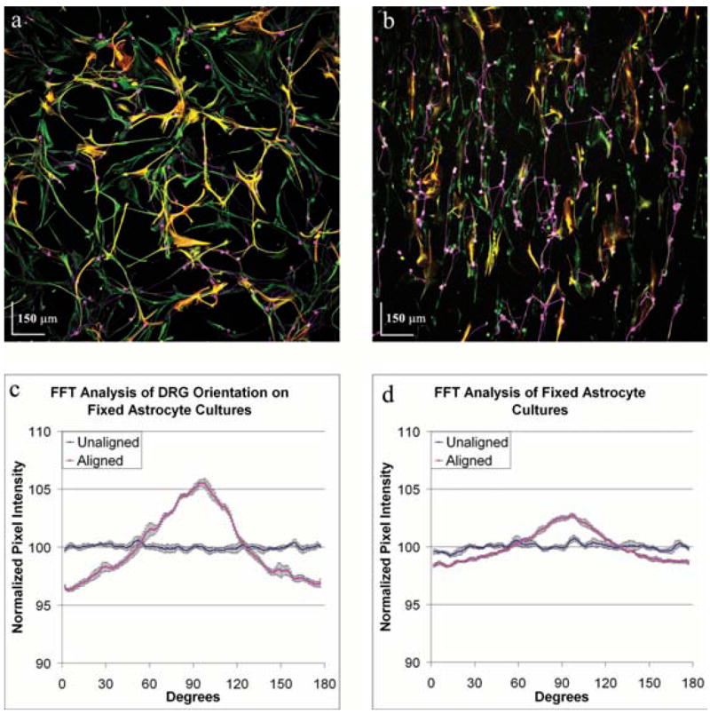
The extension and directionality of neurite outgrowth are key to achieving successful target connections during both CNS development and during the re-establishment of connections lost after neural trauma. The degree of axonal elongation depends, in large part, on the spatial arrangement of astrocytic processes rich in growth-promoting proteins. Because astrocytes in culture align their processes on exposure to an electrical field of physiological strength, we sought to determine the extent to which aligned astrocytes affect neurite outgrowth. To this end, dorsal root ganglia cells were seeded onto cultured rat astrocytes that were pre-aligned by exposure to an electric field of physiological strength (500 mV mm(-1)). Using confocal microscopy and digital image analysis, we found that neurite outgrowth at 24 hours and at 48 hours is enhanced significantly and directed consistently along the aligned astrocyte processes. Moreover, this directed neurite outgrowth is maintained when grown on fixed, aligned astrocytes. Collectively, these results indicate that endogenous electric fields present within the developing CNS might act to align astrocyte processes, which can promote and direct neurite growth. Furthermore, these results demonstrate a simple method to produce an aligned cellular substrate, which might be used to direct regenerating neurites.

Keywords: Astrocytes; DRG; alignment; electric; neurite.


Figures

Fig. 1
Fig. 1. Schematic of the electrical field chamber
Two 24-well culture plates are modified to expose simultaneously three chambers of astrocytes to an electric field. Current generated by a power supply is passed through the chamber series in the direction indicated by the arrows.
Fig. 2
Fig. 2. Electrically-aligned astrocytes guide neurite outgrowth
Astrocyte cultures were either exposed to an electric field (500 mV mm−1) for 24 hours or left unexposed as controls. A suspension of dorsal root ganglia cells was then seeded onto the astrocyte cultures and allowed to grow for 48 hours without an electrical field. Images derived from the same field of view show the spatial relationship between control astrocytes (a) and dorsal root ganglion cells (g) and between exposed astrocytes (d) and the dorsal root ganglion cells (j). Plus (+) and minus(−) signs indicate the direction of the electric field vector. (a) Control astrocytes expressing the intermediate filament Vimentin (green), appeared to be randomly oriented. FFT analysis of 12 images provided information about gross image orientation in the form of pixel intensity (b, e, h, k). The pixel intensity of the FFT image is summed along a straight line radiating at the angle, from the center to the edge of the image. (c) Plotting the normalized summed pixel intensity shows equal orientation in every direction. (d) On exposure to an electric field astrocytes display a strong alignment perpendicular to the field. (e,f) FFT analysis of six astrocyte images reveals strong orientation towards 88.8°. (g–i) Similarly, dorsal root ganglia cells seeded on control astrocytes (g) show random orientation that is identical to the astrocyte cultures (h,i). (j–l) Conversely, DRGs seeded on electrically aligned astrocytes (j) preferentially align towards 88.8° (k,l).
Fig. 3
Fig. 3. Neurites follow the processes of electrically-aligned astrocytes
Astrocyte cultures were either exposed to a electric field (500 mV mm−1) for 24 hours or left unexposed as a control. A dorsal root ganglia cell suspension was then seeded onto the astrocyte cultures and allowed to grow for 48 hours. (a,c) Cocultures of neurons (TUJ1+, magenta) and astrocytes (Vimentin+, green and GFAP+, red, which are yellow when colocalized) of control and electrically aligned cultures, respectively. + and − indicate the direction of the electric-field vector. (b,d) Higher magnification indicates that direct contact between neuronal processes and astrocytes is established. Five XZ sections (location marked 1–5) show direct contact between neurites and astrocyte processes in all three dimensions.
Fig. 4
Fig. 4. Electrically-aligned astrocytes guide neurite growth and increase neurite length
Composite images of multiple neurons grown on either control (a) or electrically aligned (c) astrocytes demonstrate the influence of astrocyte orientation on neurite outgrowth. By superimposing 37 neurons grown on unaligned, control astrocytes it is apparent that neurite outgrowth is random (a). (b) Graphically illustrating the direction of neurite outgrowth using bin sizes of 10° it is apparent that DRGs have no preferred direction of growth on randomly orientated astrocytes. The directionality coefficient for each neurite indicates an average directionality of 0.59 ± 0.03, which is consistent with randomness. (c) Superimposing the outgrowth of 39 neurons grown on electrically aligned astrocytes indicates that neurite outgrowth is aligned. (d) This is illustrated graphically by binning the directionality of neurite outgrowth. The directionality coefficient, 0.89 ± 0.02, is significantly different (P<0.001) to controls, indicating significant orientation perpendicular to the electric field direction. (e) In addition, neurite length on electrically aligned astrocytes is significantly greater (P<0.001, *) than on unaligned astrocytes.
Fig. 5
Fig. 5. Continued exposure to electric fields enhances neurite and astrocyte alignment
Astrocyte alignment following cessation of electric-field exposure was maintained by exposing aligned astrocytes to an additional weak (10 mV mm−1) or strong (500 mV mm−1) electric field for 24 hours. Cells were stained as described in Fig. 2. (a) FFT analysis of these cultures demonstrates that astrocyte alignment is improved by exposure to a continued electric field of 500 mV mm−1. The influence of continued exposure to an electric field and the enhanced alignment of astrocytes on neurite outgrowth was examined by seeding dorsal root ganglia cells onto aligned astrocytes and exposing the co-culture to 500 mV mm−1 for 24 hours. (b) FFT analysis of neurite orientation shows that continued exposure to electric fields improves alignment. (c) The alignment was quantified by comparing the volume under the curve within ±10° of the peak, which showed a significant increase in alignment of neurites exposed to 500 mV mm−1 (P<0.01, *, P<0.01, **). Examination of neurite length, as in Fig. 3, shows that the total length of neurites is significantly longer when grown on electrically aligned astrocytes compared with unaligned astrocytes (P<0.01, *). (d) With continued exposure to an electric field, there is no longer an increase in neurite length.
Fig. 6
Fig. 6. Fixation of aligned astrocytes provides a substrate that induces neurite alignment
Astrocyte cultures were aligned by exposure to an electric field of 500 mV mm−1 for 24 hours and then fixed immediately in paraformaldehyde. Cells were stained as described in Fig. 2. The astrocyte cultures were washed with PBS and dorsal root ganglia cells were seeded onto the cultures and allowed to grow for 24 hours. FFT analysis of six images of DRGs grown on fixed unaligned astrocytes (a) and six images of DRGs grown on fixed electrically-aligned astrocyte cultures (b) show that the direction of neurite growth (c) is in the direction of astrocyte alignment on the fixed electrically aligned astrocytes (d, red line), whereas orientation of neurites is random on the unaligned astrocytes (d, blue line).

Similar articles

Cited by

References

    1. Bandtlow CE, Schwab ME. NI-35/250/nogo-a: a neurite growth inhibitor restricting structural plasticity and regeneration of nerve fibers in the adult vertebrate CNS. Glia. 2000;29:175–181. - PubMed
    1. Biran R, Noble MD, Tresco PA. Directed nerve outgrowth is enhanced by engineered glial substrates. Experimental Neurology. 2003;184:141–152. - PubMed
    1. Borgens RB, Shi R, Mohr TJ, Jaeger CB. Mammalian cortical astrocytes align themselves in a physiological voltage gradient. Experimental Neurology. 1994;128:41–49. - PubMed
    1. Borgens RB, Vanable JW, Jr, Jaffe LF. Reduction of sodium dependent stump currents disturbs urodele limb regeneration. Journal of Experimental Zoology. 1979;209:377–386. - PubMed
    1. Cameron RS, Rakic P. Glial cell lineage in the cerebral cortex: a review and synthesis. Glia. 1991;4:124–137. - PubMed

LinkOut - more resources