. 2008:6854:68540H.
doi: 10.1117/12.768798.

Prosthetic systems for therapeutic optical activation and silencing of genetically-targeted neurons

Affiliations

Prosthetic systems for therapeutic optical activation and silencing of genetically-targeted neurons

Jacob G Bernstein et al. Proc SPIE Int Soc Opt Eng. 2008.

Abstract

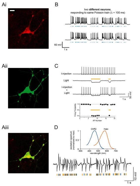
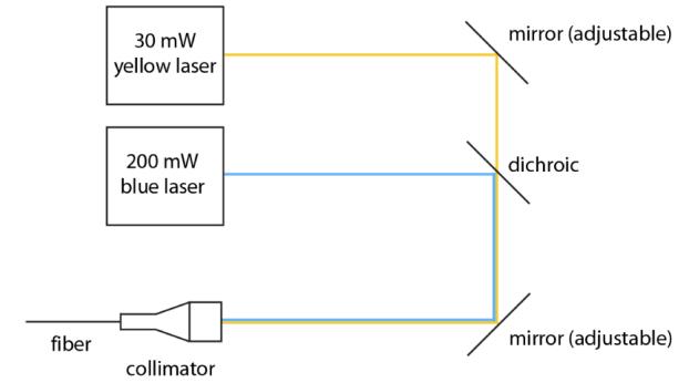
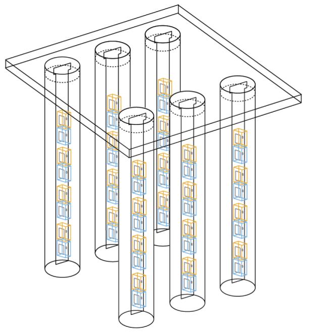
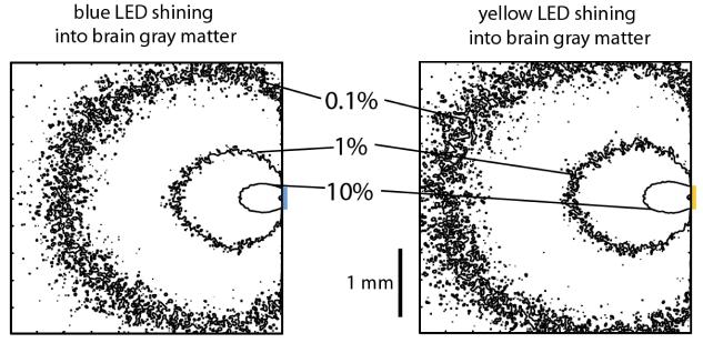
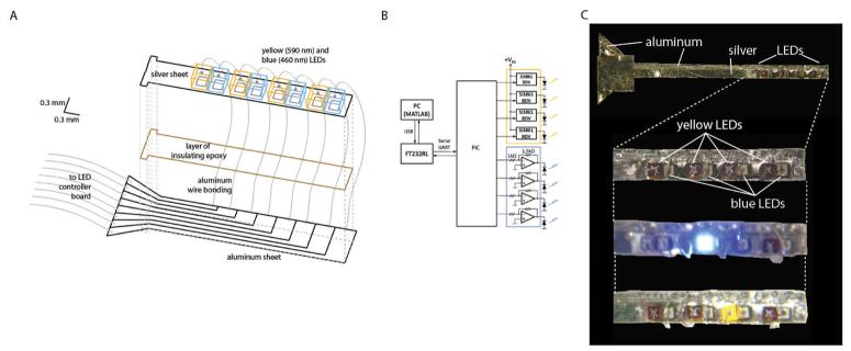
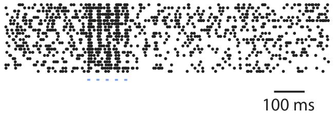
Many neural disorders are associated with aberrant activity in specific cell types or neural projection pathways embedded within the densely-wired, heterogeneous matter of the brain. An ideal therapy would permit correction of activity just in specific target neurons, while leaving other neurons unaltered. Recently our lab revealed that the naturally-occurring light-activated proteins channelrhodopsin-2 (ChR2) and halorhodopsin (Halo/NpHR) can, when genetically expressed in neurons, enable them to be safely, precisely, and reversibly activated and silenced by pulses of blue and yellow light, respectively. We here describe the ability to make specific neurons in the brain light-sensitive, using a viral approach. We also reveal the design and construction of a scalable, fully-implantable optical prosthetic capable of delivering light of appropriate intensity and wavelength to targeted neurons at arbitrary 3-D locations within the brain, enabling activation and silencing of specific neuron types at multiple locations. Finally, we demonstrate control of neural activity in the cortex of the non-human primate, a key step in the translation of such technology for human clinical use. Systems for optical targeting of specific neural circuit elements may enable a new generation of high-precision therapies for brain disorders.


Figures

Fig. 1
Fig. 1
A, Neuron expressing channelrhodopsin-2 fused to mCherry (Ai; bar, 20 [μm) and halorhodopsin fused to GFP (Aii); overlay shown in Aiii. B, Poisson trains of spikes elicited by pulses of blue light (blue dashes), in two different neurons. C, Light-driven spike blockade, demonstrated (Top) for a representative hippocampal neuron, and (Bottom) for a population of neurons (n = 7). I-injection, neuronal firing induced by pulsed somatic current injection (300 pA, 4 ms). Light, hyperpolarization induced by periods of yellow light (yellow dashes). I-injection + Light, yellow light drives Halo to block neuron spiking, leaving spikes elicited during periods of darkness intact. D, Top, action spectrum for ChR2 overlaid with absorption spectrum for N. pharaonis halorhodopsin. Bottom, Hyperpolarization and depolarization events induced in a representative neuron by a Poisson train of alternating pulses (10 ms) of yellow and blue light. Adapted from and .
Fig. 2
Fig. 2
Neurons in the mouse brain expressing Halo-GFP under the CaMKII promoter, which preferentially labels excitatory neurons. Scalebar, 50 μm.
Fig. 3
Fig. 3
Schematic of a setup that allows blue and yellow light to be focused down a fiber to be implanted inside the head, for bi-directional control of a single kind of neuron at a single site within the brain, or for activation and silencing of two different kinds of neuron at a single site within the brain.
Fig. 4
Fig. 4
Schematic of a setup that enables 3-D arrays of yellow and blue LEDs to activate, silence, and resculpt neural activity in arbitrary 3-D patterns. The device is a 2-D array made of 1-D LED-bearing probes, each of which has alternating blue and yellow LEDs going down its length. The 1-D probes are placed in glass capillaries to prevent brain heating. The capillaries are then arranged in a 2-D array by placing them in form-fitting holes within a plastic plate.
Fig. 5
Fig. 5
Monte Carlo simulations of how blue (left) and yellow (right) photons travel through the brain from LEDs (represented by colored lines to the right of the square). Shown are squares representing 4 mm × 4 mm cross-sections taken through the 4 mm × 4 mm × 4 mm cube simulated; the cross-sections are taken by slicing the cube through the LED center. Contours represent cross-sections of the surfaces at which the radiant flux drops to 10%, 1%, or 0.1% of their values on the LED surface.
Fig. 6
Fig. 6
A, schematic of a 1-D LED probe, the basic building block of the 3-D array schematized in Fig. 4, expanded for ease of seeing how the probe is constructed. B, LED driver circuit, made of op-amps, load drivers, and a microcontroller. C, From top to bottom: picture of whole 1-D LED probe, zoomed-in picture of the LED part of the probe, zoomed-in picture of the LED part of the probe with one blue LED on, zoomed-in picture of the LED part of the probe with one yellow LED on.
Fig. 7
Fig. 7
Raster plots indicate occurrences of spikes (black dots) elicited from an excitatory neuron expressing ChR2 in the monkey cortex, in 22 consecutive spike trains (each shown as a row of black dots) elicited in response to a brief train of blue light stimulation. Each horizontal row reflects one recording of the response to five blue pulses of light (shown as blue dashes), each lasting 10 ms and separated from the next by 20 ms (i.e., 33.3 Hz stimulation rate).

References

    1. Bernard C, Anderson A, Becker A, et al. Science. 2004;305(5683):532. - PubMed
    2. Sanabria ER, Su H, Yaari Y. J Physiol. 2001;532(Pt 1):205. - PMC - PubMed
    3. Shao LR, Dudek FE. J Neurophysiol. 2004;92(3):1366. - PubMed
    4. Houser CR, Miyashiro JE, Swartz BE, et al. J Neurosci. 1990;10(1):267. - PMC - PubMed
    1. Buckmaster PS, Dudek FE. J Comp Neurol. 1997;385(3):385. - PubMed
    1. Lewis DA, Hashimoto T, Volk DW. Nat Rev Neurosci. 2005;6(4):312. - PubMed
    1. George MS, Wassermann EM, Williams WA, et al. Neuroreport. 1995;6(14):1853. - PubMed
    2. Pascual-Leone A, Rubio B, Pallardo F, et al. Lancet. 1996;348(9022):233. - PubMed
    3. Mayberg HS, Lozano AM, Voon V, et al. Neuron. 2005;45(5):651. - PubMed
    1. Groves DA, Brown VJ. Neuroscience and biobehavioral reviews. 2005;29(3):493. - PubMed
    2. Fountas KN, Smith JR, Murro AM, et al. Stereotactic and functional neurosurgery. 2005;83(4):153. - PubMed
    3. Morrell M. Current opinion in neurology. 2006;19(2):164. - PubMed

LinkOut - more resources