. 2008 Jul;189(1):35-47.
doi: 10.1007/s00221-008-1401-1. Epub 2008 May 6.

Coordination of eye and head components of movements evoked by stimulation of the paramedian pontine reticular formation

Affiliations

Coordination of eye and head components of movements evoked by stimulation of the paramedian pontine reticular formation

Neeraj J Gandhi et al. Exp Brain Res. 2008 Jul.

Abstract

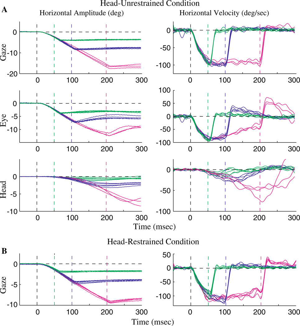
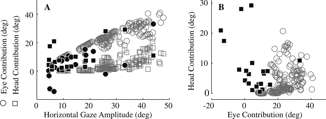
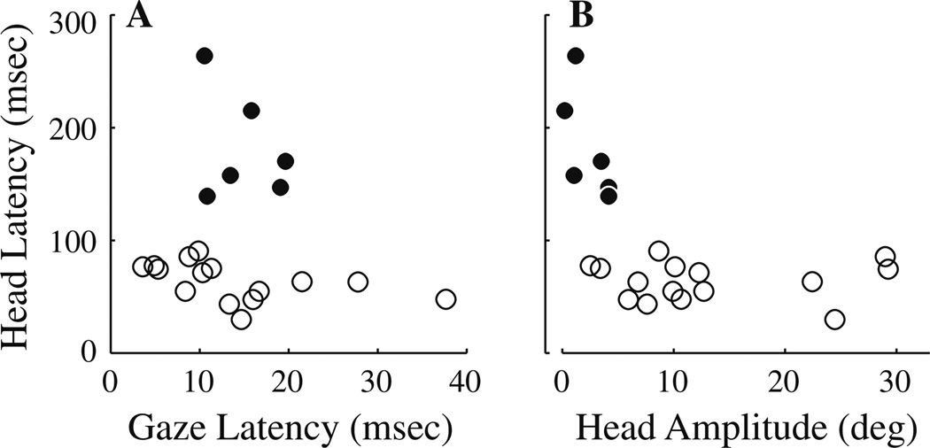
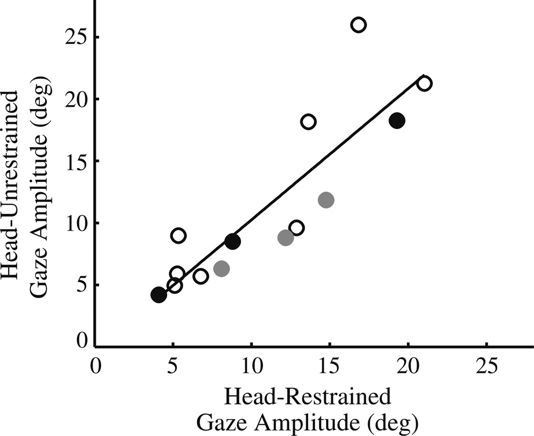
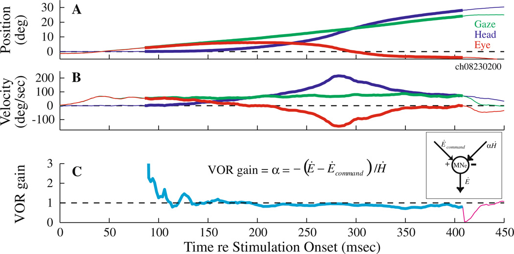
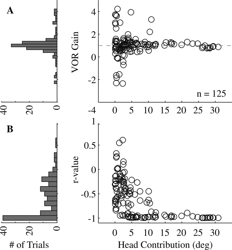
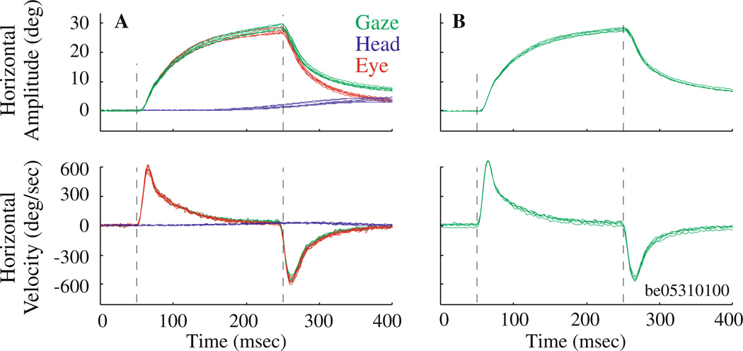
Constant frequency microstimulation of the paramedian pontine reticular formation (PPRF) in head-restrained monkeys evokes a constant velocity eye movement. Since the PPRF receives significant projections from structures that control coordinated eye-head movements, we asked whether stimulation of the pontine reticular formation in the head-unrestrained animal generates a combined eye-head movement or only an eye movement. Microstimulation of most sites yielded a constant-velocity gaze shift executed as a coordinated eye-head movement, although eye-only movements were evoked from some sites. The eye and head contributions to the stimulation-evoked movements varied across stimulation sites and were drastically different from the lawful relationship observed for visually-guided gaze shifts. These results indicate that the microstimulation activated elements that issued movement commands to the extraocular and, for most sites, neck motoneurons. In addition, the stimulation-evoked changes in gaze were similar in the head-restrained and head-unrestrained conditions despite the assortment of eye and head contributions, suggesting that the vestibulo-ocular reflex (VOR) gain must be near unity during the coordinated eye-head movements evoked by stimulation of the PPRF. These findings contrast the attenuation of VOR gain associated with visually-guided gaze shifts and suggest that the vestibulo-ocular pathway processes volitional and PPRF stimulation-evoked gaze shifts differently.


Figures

Fig. 1
Fig. 1
Effects of microstimulation of the PPRF region in monkey. a–d Stimulation delivered with the head immobilized. Each panel superimposes several traces (n = 4–5) of horizontal amplitude (top) and velocity (bottom) waveforms of stimulation-evoked movements from four different sites. Stimulation parameters: a 25 µA, 200 ms, 300 Hz; b 20 µA, 400 ms, 300 Hz; c 25 µA, 400 ms, 300 Hz; d 20 µA, 400 ms, 400 Hz. Vertical dashed lines indicate stimulation onset and offset, and horizontal dashed lines mark zero displacement and velocity. The initial position was subtracted from each trace to align the movements. e–h Stimulation delivered with the head unrestrained. Each panel superimposes representative stimulation-evoked movements (n = 3–7) evoked from the same four sites and using the same parameters as above. Top and bottom panels plot horizontal components of representative stimulation-evoked changes in amplitude and velocity, respectively, of gaze (green), head-in-space (blue), and eye-in-head (red)
Fig. 2
Fig. 2
Effects of varying stimulation duration on eye-head movements evoked by stimulation of the PPRF region. a Head-unrestrained condition. The green, blue, and magenta traces show movements when stimulation duration was set to 50, 100, and 200 ms, respectively. Stimulation intensity (25 µA) and frequency (300 Hz) were held constant. The individual rows plot horizontal gaze, eye-in-head and head amplitude (left column) and velocity (right column) for representative, individual trials. The first vertical line marks stimulation onset, and the remaining vertical lines denote stimulation offset for the three different durations (indicated by the different colors). b Head-restrained condition. The same format as above, but only the gaze channel is shown as the head was held stationary. Thus, the eye component equals the gaze component
Fig. 3
Fig. 3
Distribution of eye and head contributions. a Eye (○) and head (□) contributions are plotted as a function of horizontal gaze amplitude evoked by stimulation of the PPRF region (filled symbols; n = 21 sites) and for visually-guided, horizontal gaze shifts (open, gray symbols; n = 200). Note that eye contributions can be negative for positive, stimulation-evoked changes in gaze (e.g., Fig. 1f, g). For the visually-guided gaze shifts, the initial eye positions were limited to ±5° and initial head positions were restricted to ±10°. For the stimulation-evoked movements, the initial eye positions were limited to ±12° and initial head positions were restricted to ±10°. b An alternate representation of panel a. Head contribution is plotted as function of eye contribution for visually-guided gaze shifts (○) and for eye-head movement evoked by stimulation of the PPRF (■). The distributions of two types of movements are clearly different
Fig. 4
Fig. 4
Analyses of the latency of stimulation-evoked movements. a Head latency is plotted against gaze latency. Apart from the six sites for which head latency was prolonged (●), head onset followed gaze onset by 49.6 ± 22.9 ms. b Plotting head latency against head amplitude shows that these six sites (●) were associated with sites with minimal head movements, rendering detection of head onset problematic
Fig. 5
Fig. 5
Comparison of stimulation-evoked changes in gaze in head-restrained and head-unrestrained conditions. The data are fitted with a linear regression (solid line; slope = 1.05; r = 0.87; P< 0.0001). Data are from 10 sites. The open circles indicate the 8 sites with only one configuration of stimulation parameters. For two sites (black and gray circles), data points for each of three sets of stimulation parameters are included. For the site shown in filled gray circles, the stimulation durations were 200, 300, and 400 ms (20 µA, 300 Hz). For the site represented with filled black circles (see Fig. 2), stimulation durations were 50, 100, and 200 ms (25 µA, 300 Hz)
Fig. 6
Fig. 6
Example illustration of VOR gain computation during coordinated eye-head movements generated by PPRF stimulation. Temporal traces of the horizontal position (a) and velocity (b) of gaze (green), head (blue) and eye-in-head (red) components are shown for one trial. Plot starts at stimulation onset. The thick overlays on the traces mark the period from head movement onset to the end of the stimulation train. c The instantaneous VOR gain (α = − (Ė − Ėcommand)/, see text for details) is plotted as a function of time. During the stimulation epoch (cyan trace), the velocity command (Ėcommand) equaled the constant velocity observed in the head-restrained condition at the same site and with identical stimulation parameters. It was set to zero after stimulation offset (magenta trace). The inset shows a schematic in which the output of the extraocular motoneurons incorporates an excitatory velocity input from the PPRF (saccade pathway) and an inhibitory velocity input due to the head movement (vestibular pathway)
Fig. 7
Fig. 7
Results of the linear regression fits. a The estimated VOR gain is plotted in histogram format (left) and as a function of head contribution (right). The head contribution equals the displacement of the head during the change in gaze evoked by PPRF stimulation. The analysis was performed for the 125 trials across 10 stimulation sites. The dashed horizontal line indicates unity VOR gain. b The correlation coefficient or, the r value, of each individual linear fit is shown in the same format as a
Fig. 8
Fig. 8
Microstimulation of the abducens nucleus in a head-unrestrained and b head-restrained conditions. a Top and bottom panels plot horizontal components of stimulation-evoked changes in position and velocity, respectively, in gaze (green), eye-in-head (red) and head-in-space (blue). b Same format at a, but only gaze profiles (green) are displayed; eye-in-head equals gaze because head-in-space is held constant at zero. Stimulation parameters: 25 µA, 200 ms, 300 Hz. Vertical dashed lines indicate stimulation onset and offset. These representative trials for both head-restrained (n = 5 trials) and head-unrestrained (n = 7 trials) data were evoked from the same site

Similar articles

Cited by

References

    1. Andre-Deshays C, Berthoz A, Revel M. Eye-head coupling in humans. I. Simultaneous recording of isolated motor units in dorsal neck muscles and horizontal eye movements. Exp Brain Res. 1988;69:399–406. - PubMed
    1. Breznen B, Lu SM, Gnadt JW. Analysis of the step response of the saccadic feedback: system behavior. Exp Brain Res. 1996;111:337–344. - PubMed
    1. Chen LL, Walton MM. Head movement evoked by electrical stimulation in the supplementary eye field of the rhesus monkey. J Neurophysiol. 2005;94:4502–4519. - PubMed
    1. Cohen B, Komatsuzaki A. Eye movements induced by stimulation of the pontine reticular formation: evidence for integration in oculomotor pathways. Exp Neurol. 1972;36:101–117. - PubMed
    1. Corneil BD, Olivier E, Munoz DP. Neck muscle activity evoked by stimulation of the monkey superior colliculus. I. Topography and manipulation of stimulation parameters. J Neurophysiol. 2002;88:1980–1999. - PubMed

Publication types

MeSH terms

LinkOut - more resources