Comparative Study
. 2009 May;24(3):313-20.
doi: 10.1007/s10103-008-0561-3. Epub 2008 May 6.

Decontamination efficacy of erbium:yttrium-aluminium-garnet and diode laser light on oral Candida albicans isolates of a 5-day in vitro biofilm model

Affiliations
Comparative Study

Decontamination efficacy of erbium:yttrium-aluminium-garnet and diode laser light on oral Candida albicans isolates of a 5-day in vitro biofilm model

Sabine Sennhenn-Kirchner et al. Lasers Med Sci. 2009 May.

Abstract

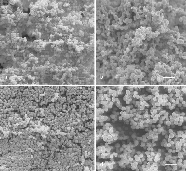
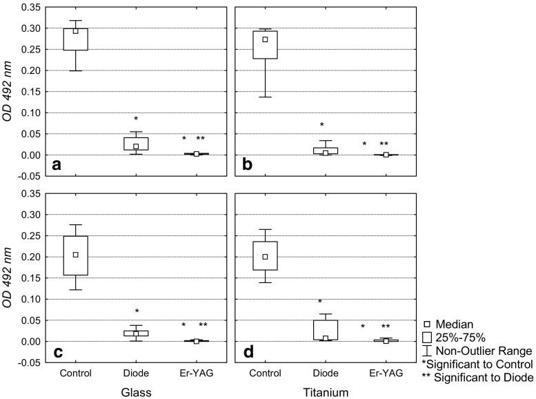
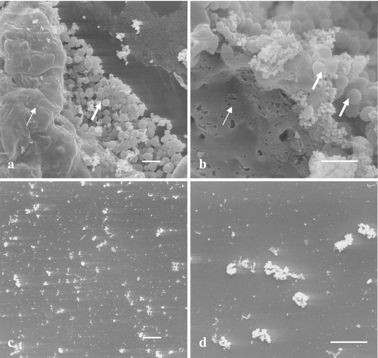
The different forms of superficial and systemic candidiasis are often associated with biofilm formation on surfaces of host tissues or medical devices. The biofilm formation of Candida spp., in general, necessitates significantly increased amounts of antifungal agents for therapy. Often the therapeutic effect is doubtful. A 5-day biofilm model with oral Candida isolates was established according to Chandra et al. (J Dent Res 80:903-908, 2001) on glass and titanium surfaces and was modified by Sennhenn-Kirchner et al. (Z Zahnärztl Implantol 3:45-51, 2007) to investigate different aspects unanswered in the field of dentistry. In this model, the efficacy of erbium:yttrium-aluminium-garnet (Er:YAG) light (2940 nm, 100 mJ, 10 Hz, 300 micros pulsed mode applied for 80 s) and diode laser light (810 nm, 1 W, continuous wave mode applied for 20 s with four repetitions after 30 s pauses each) was evaluated and compared to untreated controls. The photometric evaluation of the samples was completed by observations on morphological changes of yeast cells grown in the biofilm. Compared to the untreated controls Candida cells grown in mature in vitro biofilms were significantly reduced by both wavelengths investigated. Comparison between the different methods of laser treatment additionally revealed a significantly greater effect of the Er:YAG over the diode laser. Scanning electron microscopy findings proved that the diode laser light was effective in direct contact mode. In contrast, in the areas without direct contact, the fungal cells were left almost unchanged. The Er:YAG laser damaged the fungal cells to a great extent wherever it was applied.


Figures

Fig. 1
Fig. 1
SEM image of Candida albicans biofilms grown for 5 days on glass and titanium surfaces. No morphological difference between the two isolates C. alb. SK1 (a, c) and C. alb. SK2 (b, d) colonising glass (a, b) and titanium (c, d) surfaces became obvious in the SEM evaluation. Blastoconidia and pseudomycelia could be observed. All ×1,000. The bars represent 10 μm
Fig. 2
Fig. 2
Box plots of the efficacy of the treatment of the Candida biofilms by diode and Er:YAG irradiation on glass and titanium surfaces. The results of the evaluation on the oral isolates C. albicans SK1 (a and b) and C. albicans SK 2 (c and d) are depicted, in comparison with the control values. The trial was performed at least in duplicate with six repetitions. The absolute values (OD 492 nm) are depicted on the ordinates. The reduction in the viability of Candida albicans cells following the irradiation by diode and Er:YAG laser light for 80 s each is shown. Compared to the controls, the reduction with both laser regimens was statistically significant (*). A statistically significant difference was also demonstrated for the comparison of diode and Er:YAG laser (**). OD optical density
Fig. 3
Fig. 3
Efficacy of the diode (a, b) and the Er:YAG (c, d) laser treatments demonstrated by SEM. a, c ×1,000; b, d ×3,000. The bars represent 10 μm. The Candida cells seem to be squashed and melted due to the direct contact with the laser fibre (thin arrows). In other areas the fungal cells are left almost unchanged (thick arrows). In combination with the cooling water, this Er:YAG laser treatment damaged the fungal cells to a greater extent wherever it reached the surface and nearly removed the damaged cells from the surface of the slides

Similar articles

Cited by

References

    1. Kojic EM, Darouiche RO. Candida infections of medical devices. Clin Microbiol Rev. 2004;17:255–267. doi: 10.1128/CMR.17.2.255-267.2004. - DOI - PMC - PubMed
    1. Ramage G, Tomsett K, Wickes BL, López-Ribot JL, Redding SW. Denture stomatitis: a role for Candida biofilms. Oral Surg Oral Med Oral Pathol Oral Radiol Endod. 2004;98:53–59. doi: 10.1016/j.tripleo.2003.04.002. - DOI - PubMed
    1. Jarvis WR. Epidemiology of nosocomial fungal infections, with emphasis on Candida species. Clin Infect Dis. 1995;20:1526–1530. doi: 10.1093/clinids/20.6.1526. - DOI - PubMed
    1. Ramage G, Saville SP, Thomas DP, López-Ribot JL. Candida biofilms: an update. Eukaryot Cell. 2005;4:633–638. doi: 10.1128/EC.4.4.633-638.2005. - DOI - PMC - PubMed
    1. Sandven P. Epidemiology of candidemia. Rev Iberoam Micol. 2000;17:73–81. - PubMed

Publication types

MeSH terms

Substances

LinkOut - more resources