. 2008 Jul;466(7):1583-91.
doi: 10.1007/s11999-008-0264-x. Epub 2008 May 6.

Loss of homeostatic strain alters mechanostat "set point" of tendon cells in vitro

Affiliations

Loss of homeostatic strain alters mechanostat "set point" of tendon cells in vitro

Steven P Arnoczky et al. Clin Orthop Relat Res. 2008 Jul.

Abstract

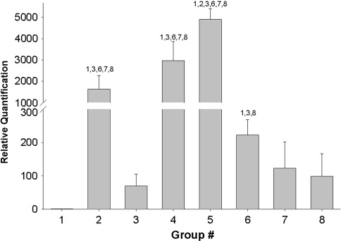
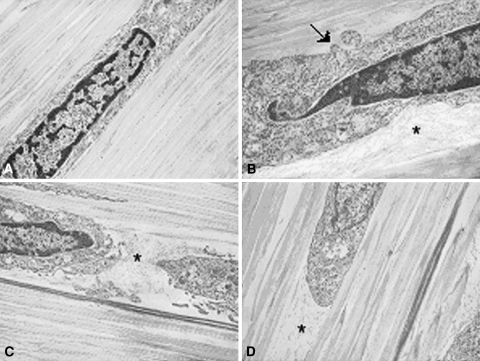
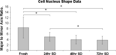
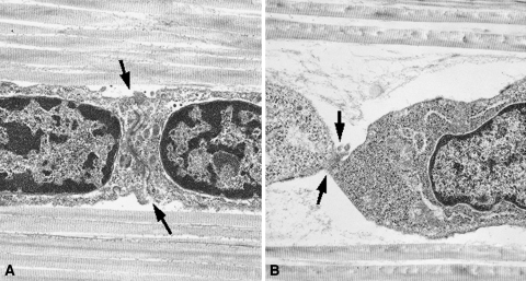
Tendon cells respond to mechanical loads. The character (anabolic or catabolic) and sensitivity of this response is determined by the mechanostat set point of the cell, which is governed by the cytoskeleton and its interaction with the extracellular matrix. To determine if loss of cytoskeletal tension following stress deprivation decreases the mechanoresponsiveness of tendon cells, we cultured rat tail tendons under stress-deprived conditions for 48 hours and then cyclically loaded them for 24 hours at 1%, 3%, or 6% strain at 0.17 Hz. Stress deprivation upregulated MMP-13 mRNA expression and caused progressive loss of cell-matrix contact compared to fresh controls. The application of 1% strain to fresh tendons for 24 hours inhibited MMP-13 mRNA expression compared to stress-deprived tendons over the same period. However, when tendons were stress-deprived for 48 hours and then subjected to the same loading regime, the inhibition of MMP-13 mRNA expression was decreased. In stress-deprived tendons, it was necessary to increase the strain magnitude to 3% to achieve the same level of MMP-13 mRNA inhibition seen in fresh tendons exercised at 1% strain. The data suggest loss of cytoskeletal tension alters the mechanostat set point and decreases the mechanoresponsiveness of tendon cells.


Figures

Fig. 1
Fig. 1
The bar graph shows the change in the inhibitory effect of cyclic tensile loading on MMP-13 mRNA expression following 48 hours of stress deprivation. All experimental samples were quantified relative to the 0 hour fresh control. Group 1: fresh (0 hour control); Group 2: stress-deprived for 24 hours; Group 3: 1% cyclic strain at 0.17 Hz for 24 hours; Group 4: stress-deprived for 48 hours; Group 5: stress-deprived for 72 hours; Group 6: stress-deprived for 48 hours followed by 1% cyclic strain at 0.17 Hz for 24 hours; Group 7: stress-deprived for 48 hours followed by 3% cyclic strain at 0.17 Hz for 24 hours; Group 8: stress-deprived for 48 hours followed by 6% cyclic strain at 0.17 Hz for 24 hours. Numbers above each bar signify significant (p < 0.05) differences between specific groups.
Fig. 2A–D
Fig. 2A–D
Transmission electron photomicrographs of (A) fresh, and (B) 24-hour, (C) 48-hour, and (D) 72-hour stress-deprived rat tail tendons demonstrating a progressive loss of contact between the cells and the extracellular matrix (ECM). Fine, fibrillar ECM components become prominent in the pericellular space (asterisk) and the normally uniform cell borders developed irregular protrusions (arrow). Collagen Type I fibril packing adjacent to the cells also appears less dense. (Stain, osmium tetroxide, uranyl acetate, and lead citrate; original magnification, ×10,000).
Fig. 3
Fig. 3
The bar graph shows the decrease in cell nuclear aspect ratios seen in stress-deprived tendons as a function of time. The decrease in the ratio of major axis length to minor axis length reflects the “rounding” up of the cells in response to the loss of homeostatic strain. The symbol (*) signifies significant (p < 0.05) differences between specific groups.
Fig. 4A–B
Fig. 4A–B
Transmission electron photomicrographs demonstrating representative cell-to-cell junctions (arrows) from (A) fresh and (B) a 48-hour stress-deprived rat tail tendon. The decrease in cell-to-cell contact area in the stress-deprived tendon could affect the extent of gap junctional intercellular communications. Loose, fine, fibrillar material is visible in the pericellular space. (Stain, osmium tetroxide, uranyl acetate, and lead citrate; original magnification, ×10,000).

References

    1. {'text': '', 'ref_index': 1, 'ids': [{'type': 'DOI', 'value': '10.1016/S0736-0266(03)00107-4', 'is_inner': False, 'url': 'https://doi.org/10.1016/s0736-0266(03)00107-4'}, {'type': 'PubMed', 'value': '14554207', 'is_inner': True, 'url': 'https://pubmed.ncbi.nlm.nih.gov/14554207/'}]}
    2. Alfredson H, Lorentzon M, Bäckman S, Bäckman A, Lerner UH. cDNA arrays and real time qualitative PCR techniques in the investigation of chronic Achilles tendinosis. J Orthop Res. 2003;21:970–975. - PubMed
    1. None
    2. Arnoczky SP, Lavagnino M, Egerbacher M. The response of tendon cells to changing loads: implications in the etiopathogenesis of tendinopathy. In: Woo S-L-Y, Renstrom P, Arnoczky SP, eds. Understanding and Prevention of Tendinopathy in the Athlete, Encyclopedia of Sports Medicine. Blackwell Publishing: Oxford, England; 2007:46–59.
    1. {'text': '', 'ref_index': 1, 'ids': [{'type': 'DOI', 'value': '10.1111/j.1365-2613.2007.00548.x', 'is_inner': False, 'url': 'https://doi.org/10.1111/j.1365-2613.2007.00548.x'}, {'type': 'PMC', 'value': 'PMC2517314', 'is_inner': False, 'url': 'https://pmc.ncbi.nlm.nih.gov/articles/PMC2517314/'}, {'type': 'PubMed', 'value': '17696902', 'is_inner': True, 'url': 'https://pubmed.ncbi.nlm.nih.gov/17696902/'}]}
    2. Arnoczky SP, Lavagnino M, Egerbacher M. The mechanobiological aetiopathogenesis of tendinopathy: is it the over-stimulation or the under-stimulation of tendon cells? Int J Exp Path. 2007;88:217–226. - PMC - PubMed
    1. {'text': '', 'ref_index': 1, 'ids': [{'type': 'DOI', 'value': '10.1016/S0736-0266(01)00080-8', 'is_inner': False, 'url': 'https://doi.org/10.1016/s0736-0266(01)00080-8'}, {'type': 'PubMed', 'value': '11853087', 'is_inner': True, 'url': 'https://pubmed.ncbi.nlm.nih.gov/11853087/'}]}
    2. Arnoczky SP, Lavagnino M, Whallon JJ, Hoonjan A. In situ cell nucleus deformation under tensile load: a morphologic analysis using confocal laser microscopy. J Orthop Res. 2002;20:29–35. - PubMed
    1. {'text': '', 'ref_index': 1, 'ids': [{'type': 'DOI', 'value': '10.1016/S0736-0266(03)00185-2', 'is_inner': False, 'url': 'https://doi.org/10.1016/s0736-0266(03)00185-2'}, {'type': 'PubMed', 'value': '15013092', 'is_inner': True, 'url': 'https://pubmed.ncbi.nlm.nih.gov/15013092/'}]}
    2. Arnoczky SP, Tian T, Lavagnino M, Gardner K. Ex vivo static tensile loading inhibits MMP-1 expression in rat-tail tendon cells through a cytoskeletally based mechanotransduction mechanism. J Orthop Res. 2004;22:328–333. - PubMed

Substances