. 2008 Aug 1;68(11):1152-64.
doi: 10.1002/pros.20786.

Expression of microRNAs and protein-coding genes associated with perineural invasion in prostate cancer

Affiliations

Expression of microRNAs and protein-coding genes associated with perineural invasion in prostate cancer

Robyn L Prueitt et al. Prostate. .

Abstract

Background: Perineural invasion (PNI) is the dominant pathway for local invasion in prostate cancer. To date, only few studies have investigated the molecular differences between prostate tumors with PNI and those without it.

Methods: To evaluate the involvement of both microRNAs and protein-coding genes in PNI, we determined their genome-wide expression with a custom microRNA microarray and Affymetrix GeneChips in 50 prostate adenocarcinomas with PNI and 7 without it. In situ hybridization (ISH) and immunohistochemistry was used to validate candidate genes.

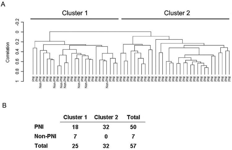
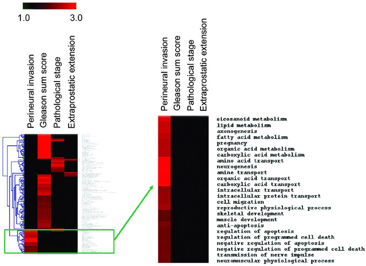
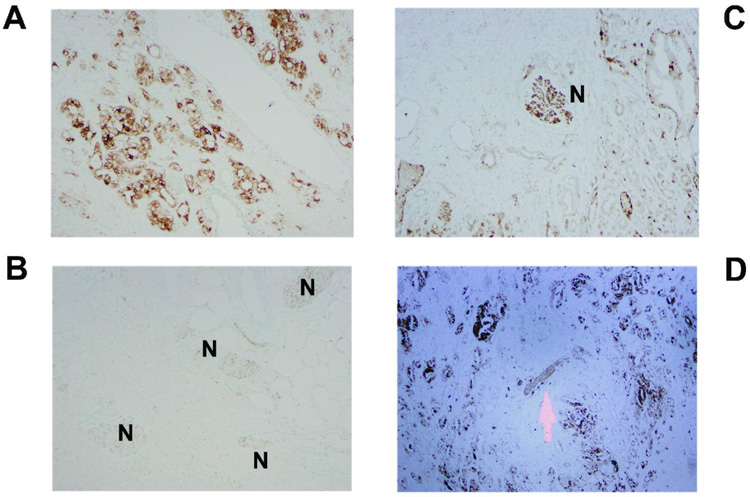
Results: Unsupervised classification of the 57 adenocarcinomas revealed two clusters of tumors with distinct global microRNA expression. One cluster contained all non-PNI tumors and a subgroup of PNI tumors. Significance analysis of microarray data yielded a list of microRNAs associated with PNI. At a false discovery rate (FDR)<10%, 19 microRNAs were higher expressed in PNI tumors than in non-PNI tumors. The most differently expressed microRNA was miR-224. ISH showed that this microRNA is expressed by perineural cancer cells. The analysis of protein-coding genes identified 34 transcripts that were differently expressed by PNI status (FDR<10%). These transcripts were down-regulated in PNI tumors. Many of those encoded metallothioneins and proteins with mitochondrial localization and involvement in cell metabolism. Consistent with the microarray data, perineural cancer cells tended to have lower metallothionein expression by immunohistochemistry than nonperineural cancer cells.

Conclusions: Although preliminary, our findings suggest that alterations in microRNA expression, mitochondrial function, and cell metabolism occur at the transition from a noninvasive prostate tumor to a tumor with PNI.


Figures

Figure 1
Figure 1
Unsupervised hierarchical cluster analysis of 57 prostate tumors based on the expression of 235 microRNAs. A: The microRNA expression yielded two prominent clusters with distinct microRNA profiles. Cluster #1 contained all non-PNI tumors. B: Non-random distribution of tumors by PNI status among the two clusters (P = 0.002; two-sided Fisher’s exact test).
Figure 2
Figure 2
Cluster analysis of Gene Ontology Biological Processes that are enriched for differently expressed genes comparing PNI tumors with non-PNI tumors. The results of a cluster analysis are displayed in a heatmap with the red color indicating an enrichment of differentially expressed genes in a biological process, e.g., eicosanoid metabolism, for a particular comparison, e.g., PNI tumor versus non-PNI tumor (“Perineural invasion”). The heatmap also shows the cluster analysis for the high (7–9) versus low (5–6) Gleason score comparison (“Gleason sum score”), the pT3 versus pT2 comparison (“Pathological stage”), and the positive versus negative extraprostatic extension comparison (“Extraprostatic extension”). Our analysis revealed that gene expression differences are non-random and create unique patterns of frequently affected biological processes for the four comparisons. The enlarged cluster shows the biological processes that are uniquely enriched for differentially expressed genes comparing PNI tumors with non-PNI tumors. Eicosanoid metabolism, lipid metabolism, and axonogenesis are also enriched for differentially expressed genes comparing pT3 versus pT2.
Figure 3
Figure 3
Expression of metallothionein in prostate tumors by immunohistochemistry. The panels show examples of metallothionein expression in the tumor epithelium. Marked cytoplasmic expression of metallothionein in cancer cells distant to neurons (A) and absence of this expression in perineural cancer cells (B) in the same tumor. The expression of metallothionein is decreased as tumor cells approach the nerve (C,D). Arrow and “N” indicate the location of the brown stained nerve trunks. Counterstain: Methyl green.
Figure 4
Figure 4
Expression of the coxsackie adenovirus receptor in prostate tumors by immunohistochemistry. The panels show examples of receptor expression in the tumor epithelium. Membranous and cytoplasmic staining for the receptor in cancer cells distant to neurons (A) and in perineural cancer cells (B) in the same tumor. The expression of the coxsackie adenovirus receptor is decreased in perineural cancer cells (C,D). N: nerve trunk. Counterstain: Methyl green.
Figure 5
Figure 5
miR-224 in prostate tumors by in-situ hybridization. Shown are representative examples of cytoplasmic expression of miR-224 in the tumor epithelium. The granular brown staining shows the presence of miR-224. Most tumors showed weak labeling for miR-224 (A). In a subset of tumors, moderate to strong miR-224 labeling was observed in perineural cancer cells (B, C, D). N = nerve trunk. Counterstain: Hematoxylin.

References

    1. Jemal A, Siegel R, Ward E, Murray T, Xu J, Thun MJ. Cancer statistics, 2007. CA Cancer J Clin. 2007;57:43–66. - PubMed
    1. Villers A, McNeal JE, Redwine EA, Freiha FS, Stamey TA. The role of perineural space invasion in the local spread of prostatic adenocarcinoma. J Urol. 1989;142:763–768. - PubMed
    1. Bostwick DG, Grignon DJ, Hammond ME, Amin MB, Cohen M, Crawford D, Gospadarowicz M, Kaplan RS, Miller DS, Montironi R, Pajak TF, Pollack A, et al. Prognostic factors in prostate cancer. College of American Pathologists Consensus Statement 1999. Arch.Pathol.Lab Med. 2000;124:995–1000. - PubMed
    1. Montironi R, Mazzucchelli R, Scarpelli M, Lopez-Beltran A, Mikuz G. Prostate carcinoma I: prognostic factors in radical prostatectomy specimens and pelvic lymph nodes. BJU.Int. 2006;97:485–491. - PubMed
    1. Montironi R, Mazzucchelli R, Scarpelli M, Lopez-Beltran A, Mikuz G, Algaba F, Boccon-Gibod L. Prostate carcinoma II: prognostic factors in prostate needle biopsies. BJU.Int. 2006;97:492–497. - PubMed

Publication types

MeSH terms