

The diphthamide modification on elongation factor-2 renders mammalian cells resistant to ricin
- PMID: 18460012
- PMCID: PMC2562939
- DOI: 10.1111/j.1462-5822.2008.01159.x
The diphthamide modification on elongation factor-2 renders mammalian cells resistant to ricin
Abstract
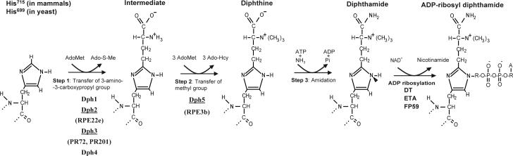
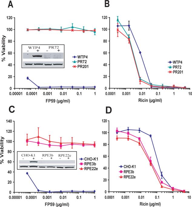
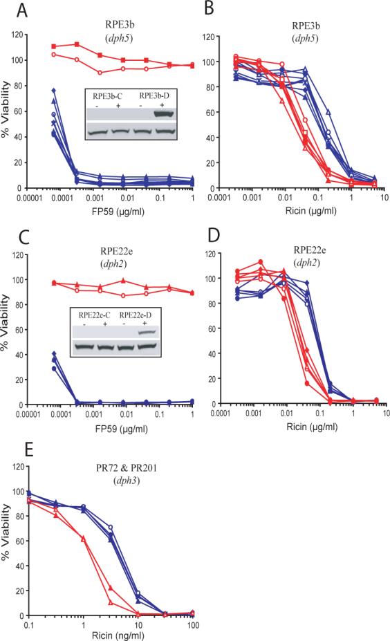
Diphthamide is a post-translational derivative of histidine in protein synthesis elongation factor-2 (eEF-2) that is present in all eukaryotes with no known normal physiological role. Five proteins Dph1-Dph5 are required for the biosynthesis of diphthamide. Chinese hamster ovary (CHO) cells mutated in the biosynthetic genes lack diphthamide and are resistant to bacterial toxins such as diphtheria toxin. We found that diphthamide-deficient cultured cells were threefold more sensitive than their parental cells towards ricin, a ribosome-inactivating protein (RIP). RIPs bind to ribosomes at the same site as eEF-2 and cleave the large ribosomal RNA, inhibiting translation and causing cell death. We hypothesized that one role of diphthamide may be to protect ribosomes, and therefore all eukaryotic life forms, from RIPs, which are widely distributed in nature. A protective role of diphthamide against ricin was further demonstrated by complementation where dph mutant CHO cells transfected with the corresponding DPH gene acquired increased resistance to ricin in comparison with the control-transfected cells, and resembled the parental CHO cells in their response to the toxin. These data show that the presence of diphthamide in eEF-2 provides protection against ricin and suggest the hypothesis that diphthamide may have evolved to provide protection against RIPs.
Figures
Similar articles
-
Identification of the proteins required for biosynthesis of diphthamide, the target of bacterial ADP-ribosylating toxins on translation elongation factor 2.Mol Cell Biol. 2004 Nov;24(21):9487-97. doi: 10.1128/MCB.24.21.9487-9497.2004. Mol Cell Biol. 2004. PMID: 15485916 Free PMC article.
-
Loss of diphthamide pre-activates NF-κB and death receptor pathways and renders MCF7 cells hypersensitive to tumor necrosis factor.Proc Natl Acad Sci U S A. 2015 Aug 25;112(34):10732-7. doi: 10.1073/pnas.1512863112. Epub 2015 Aug 10. Proc Natl Acad Sci U S A. 2015. PMID: 26261303 Free PMC article.
-
In vitro biosynthesis of diphthamide, studied with mutant Chinese hamster ovary cells resistant to diphtheria toxin.Mol Cell Biol. 1984 Apr;4(4):642-50. doi: 10.1128/mcb.4.4.642-650.1984. Mol Cell Biol. 1984. PMID: 6717439 Free PMC article.
-
The hidden nature of protein translational control by diphthamide: the secrets under the leather.J Biochem. 2019 Jan 1;165(1):1-8. doi: 10.1093/jb/mvy071. J Biochem. 2019. PMID: 30204891 Review.
-
Diphthamide - a conserved modification of eEF2 with clinical relevance.Trends Mol Med. 2024 Feb;30(2):164-177. doi: 10.1016/j.molmed.2023.11.008. Epub 2023 Dec 13. Trends Mol Med. 2024. PMID: 38097404 Review.
Cited by
-
Loss of Diphthamide Increases DNA Replication Stress in Mammalian Cells by Modulating the Translation of RRM1.ACS Cent Sci. 2024 Sep 6;10(10):1835-1847. doi: 10.1021/acscentsci.4c00967. eCollection 2024 Oct 23. ACS Cent Sci. 2024. PMID: 39463834 Free PMC article.
-
The amidation step of diphthamide biosynthesis in yeast requires DPH6, a gene identified through mining the DPH1-DPH5 interaction network.PLoS Genet. 2013;9(2):e1003334. doi: 10.1371/journal.pgen.1003334. Epub 2013 Feb 28. PLoS Genet. 2013. PMID: 23468660 Free PMC article.
-
Elongation factor 2 diphthamide is critical for translation of two IRES-dependent protein targets, XIAP and FGF2, under oxidative stress conditions.Free Radic Biol Med. 2014 Feb;67:131-8. doi: 10.1016/j.freeradbiomed.2013.10.015. Epub 2013 Oct 17. Free Radic Biol Med. 2014. PMID: 24140707 Free PMC article.
-
A structural domain mediates attachment of ethanolamine phosphoglycerol to eukaryotic elongation factor 1A in Trypanosoma brucei.PLoS One. 2010 Mar 2;5(3):e9486. doi: 10.1371/journal.pone.0009486. PLoS One. 2010. PMID: 20209157 Free PMC article.
-
A guide to taming a toxin--recombinant immunotoxins constructed from Pseudomonas exotoxin A for the treatment of cancer.FEBS J. 2011 Dec;278(23):4683-700. doi: 10.1111/j.1742-4658.2011.08182.x. Epub 2011 Jun 2. FEBS J. 2011. PMID: 21585657 Free PMC article. Review.
References
-
- Arora N, Klimpel KR, Singh Y, Leppla SH. Fusions of anthrax toxin lethal factor to the ADP-ribosylation domain of Pseudomonas exotoxin A are potent cytotoxins which are translocated to the cytosol of mammalian cells. J Biol Chem. 1992;267:15542–15548. - PubMed
-
- Barbieri L, Battelli MG, Stirpe F. Ribosome-inactivating proteins from plants. Biochim Biophys Acta. 1993;1154:237–282. - PubMed
-
- Bodley JW, Dunlop PC, VanNess BG. Diphthamide in elongation factor 2: ADP-ribosylation, purification, and properties. Methods Enzymol. 1984;106:378–387. - PubMed
Publication types
MeSH terms
Substances
Grants and funding
LinkOut - more resources
Full Text Sources
Other Literature Sources
Medical
Molecular Biology Databases
Miscellaneous
