. 2008 Jun;89(3):188-200.
doi: 10.1111/j.1365-2613.2008.00577.x.

Acute urticaria[corrected]-like lesions in allergen-unexposed cutaneous tissues in a mouse model of late allergic rhinitis

Affiliations

Acute urticaria[corrected]-like lesions in allergen-unexposed cutaneous tissues in a mouse model of late allergic rhinitis

Toshiharu Hayashi et al. Int J Exp Pathol. 2008 Jun.

Erratum in

  • Int J Exp Pathol. 2008 Aug;89(4):301

Abstract

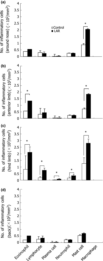
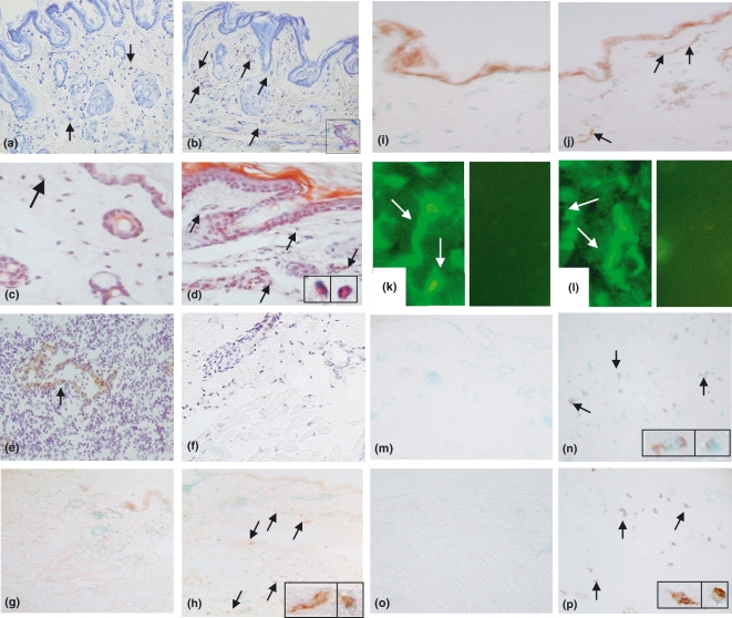
The mechanisms of distant manifestation after a local allergic reaction are largely unknown. This study examined the development of cutaneous lesions in a mouse model of late allergic rhinitis (LAR). BALB/c mice were sensitized by ovalbumin (OVA) intraperitoneally two times (on days 0 and 10) and challenged by OVA intranasally on day 14. Four days after OVA challenge, nasal and cutaneous lesions including helper T (Th) responses, expression of adhesion molecules and presence of OVA and IgE were examined, and compared with unsensitized and unchallenged (control) mice. Compared with the control group, the LAR group developed LAR characterized by infiltration of lymphocytes and eosinophils, increased IgE values and increased productions of IL-4 and IL-5, but not IFN-gamma. A dominant infiltration of eosinophils and increase in mast cells, attachment of eosinophils to endothelium, intense expression of VCAM-1 on endothelium in venules and VLA-4 expression on eosinophils and mast cells were recognized in the cutaneous tissues. There were no differences in the expression of ICAM-1 on vascular endothelium and LFA-1 on infiltrated leucocytes between the two groups. CLA expression on lymphocytes was not detected, and the binding of OVA and IgE on mast cells and eosinophils was found in the cutaneous lesions in the LAR group, but not in the control group. This study suggests that acute urticaria[corrected]-like lesions in OVA-unexposed cutaneous tissues may be induced by immediate allergic reaction due to the systemic development of Th2-type response in a mouse model of LAR.


Figures

Figure 1
Figure 1
Experimental protocol in the induction of late allergic rhinitis (LAR). On days 0 and 10, mice (n = 22) were sensitized intraperitoneally (i.p.) with 10 μg of OVA with Alum adjuvant (OVA/Alum). Four days after the second sensitization, mice were challenged intranasally (i.n.) with 500 μg of OVA (LAR group). Control group (n = 5) was injected with 100 μl of PBS i.p. on days 0 and 10, and 50 μl of PBS i.n. on day 14. Plasma was obtained on days 0 and 18 to count the number of eosinophils and measure IgE concentrations in both groups. On day 18, the whole blood, spleens, skins and the head were obtained for histopathology, immunohistochemistry and cytokine assays. Nasal (b, c, d, e and f) and cutaneous tissues (b) were sampled and examined for histopathology and immunohistochemistry (See details in Materials and methods).
Figure 2
Figure 2
Number of eosinophils (a) and IgE values (b) in blood on days 0 and 18 respectively. Each value represents the mean ± SEM. *P < 0.05.
Figure 3
Figure 3
Production of cytokines in supernatants from cultured splenocytes stimulated with Con A on day 18. Concentration of IFN-γ (a), IL-4 (b) and IL-5. Each value represents the mean ± SEM. *P < 0.05.
Figure 4
Figure 4
Evaluation of late allergic rhinitis. The thickness of nasal septum (a), number of epithelium (b), ratio of the number of goblet cell and non-goblet cell (c), score index (d; severity of inflammation) and the number of each leucocyte (e) in the respiratory region. Each value represents the mean ± SEM. *P < 0.05.
Figure 5
Figure 5
A control mouse shows relatively normal appearance with a few cell infiltration in nasal mucosa (a). Severely infiltrated eosinophils and lymphocytes with hyperplastic and degenerative respiratory epithelium are seen in a LAR mouse (b). HE ×400 (original magnification).
Figure 6
Figure 6
Number of inflammatory cells in the cutaneous tissues of the nose (a), anterior limb (b), hind limb (c) and back (d). The kinds and degrees of the infiltrating cells are variable with their location. Each value represents the mean ± SEM. *P < 0.05.
Figure 7
Figure 7
Histopathology of cutaneous tissues of the hind limb. A control mouse shows normal appearance in (a) and an arrow indicates venules (d; enlargement of a) without infiltration and destruction of vessel wall. A LAR mouse shows spongiosis with epidermal thickening (b), and infiltration of eosinophils and increase in mast cells with oedema in dermis and subcutaneous tissues (c). Details of a thin rectangle and a bold rectangle in (c) were shown in (e) and (f) respectively. (e) shows attachment of eosinophils to endothelial cells of venules (small arrows), destruction of venular wall (a bold arrow) and degranulation of mast cells (a large arrow) and (f) shows aggregation of eosinophils with degeneration and extravasation, and destruction of venular wall can be seen (a bold arrow). (g) shows peripheral nerve (an asterisk mark) with infiltration of mast cells (arrows). HE ×100 (a, b and c), ×200 (d, e, f and g) (original magnification).
Figure 8
Figure 8
Characterization of inflammatory cells and detection of adhesion molecules in cutaneous tissues of the hind limb. Inflammatory cell types, which bind to OVA and IgE, were determined by cell morphology (e.g. cell size and nuclear morphology) and by comparison with serial sections stained by HE. Representative photographs showing mast cells (a and b), eosinophils (c and d), CLA (e and f), VLA-4 (g and h), VCAM-1(i and j), ICAM-1 (k and l), OVA (m and n) and IgE (o and p) in the control (a, c, e, g, i, k, m and o) and LAR group (b, d, f, h, j, l, n and p). Degranulation of enlarged mast cell (b; insert) and degenerated eosinophils with degranulation (d; left-hand side of insert, right-hand side of insert shows node granulation of eosinophil) are visible, whereas no eosinophils are visible (c). Endothelial cells of the high endothelial venules of a lymph node (e; positive control for CLA on cutaneous lymphocytes) can be identified (an arrow), whereas no CLA-positive lymphocytes were seen (f). (h), but not (g), shows VLA-4 expression (arrows) on mast cells (h; left-hand side of insert) and eosinophils (h; right-hand side of insert). (j), but not (i), shows VCAM-1 expressions on venules (arrows). (k) and (l) show ICAM-1 expression on endothelial cells of venules (arrows), and right half of (k) and(l), which were stained without a primary antibody, shows no fluorescence. (n), but not (m), shows binding of OVA (arrows) on mast cells (n; left-hand side of insert) and eosinophils (n; right-hand side of insert). (p), but not (o), shows binding of IgE (arrows) on mast cells (p; left-hand side of insert) and eosinophils (p; right-hand side of insert). (a, b) TB; (c, d) congo red; (e–j and m–p) immunoperoxidase; (k and l) immunofluorescence. (a–j) ×100, (m–p) ×400, (k and l) ×500; inserts (b, d, h, n and p) (original magnification). Arrows indicate inflammatory cells (a-d, h, n and p) and endothelium (j, and left half of K and I).

References

    1. Beyer K, Castro R, Feidel C, Feidel C, Sampson HA. Milk-induced urticaria is associated with the expansion of T cells expressing cutaneous lymphocyte antigen. J. Allergy Clin. Immunol. 2002;109:688–693. - PubMed
    1. Brinkman L, Aslander MM, Raaijmakersb JA, Lammers JW, Koenderman L, Bruijnzeel-Koomen CA. Bronchial and cutaneous responses in atopic dermatitis patients after allergen inhalation challenge. Clin. Exp. Allergy. 1997;27:1043–1051. - PubMed
    1. Burks AW, Mallory SB, Williams LW, Shirrell MA. Atopic dermatitis: clinical relevance of food hypersensitivity reactions. J. Pediatr. 1998;113:447–451. - PubMed
    1. Cotran RS, Pober JS. Effects of cytokines on vascular endothelium: their role in vascular and immune injury. Kidney Int. 1989;35:969–975. - PubMed
    1. Franson M, Benson M, Wennergren G, Gardell LO. A role for neutrophils in intermittent allergic rhinitis. Acta Otolaryngol. 2004;124:616–620. - PubMed

Publication types

MeSH terms