

Effect of renin inhibition and AT1R blockade on myocardial remodeling in the transgenic Ren2 rat
- PMID: 18460596
- PMCID: PMC2493592
- DOI: 10.1152/ajpendo.00752.2007
Effect of renin inhibition and AT1R blockade on myocardial remodeling in the transgenic Ren2 rat
Abstract
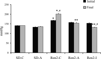
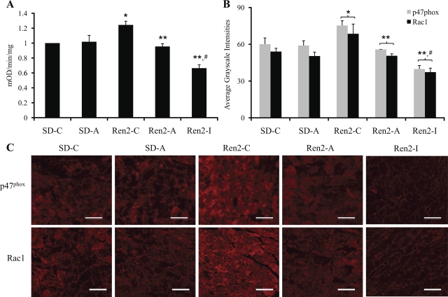
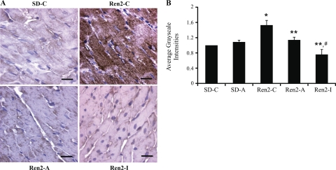
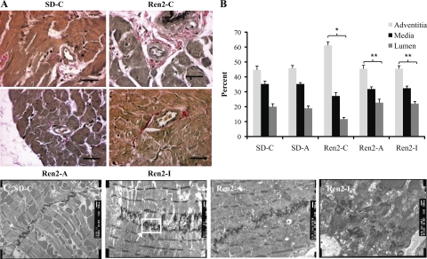
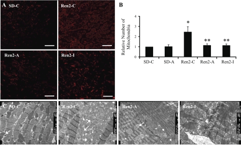
Angiotensin II (Ang II) stimulation of the Ang type 1 receptor (AT(1)R) facilitates myocardial remodeling through NADPH oxidase-mediated generation of oxidative stress. Components of the renin-angiotensin system constitute an autocrine/paracrine unit in the myocardium, including renin, which is the rate-limiting step in the generation of Ang II. This investigation sought to determine whether cardiac oxidative stress and cellular remodeling could be attenuated by in vivo renin inhibition and/or AT(1)R blockade in a rodent model of chronically elevated tissue Ang II levels, the transgenic (mRen2)27 rat (Ren2). The Ren2 overexpresses the mouse renin transgene with resultant hypertension, insulin resistance, and cardiovascular damage. Young (6- to 7-wk-old) heterozygous (+/-) male Ren2 and age-matched Sprague-Dawley rats were treated with the renin inhibitor aliskiren, which has high preferential affinity for human and mouse renin, an AT(1)R blocker, irbesartan, or placebo for 3 wk. Myocardial NADPH oxidase activity and immunostaining for NADPH oxidase subunits and 3-nitrotyrosine were evaluated and remodeling changes assessed by light and transmission electron microscopy. Blood pressure, myocardial NADPH oxidase activity and subunit immunostaining, 3-nitrotyrosine, perivascular fibrosis, mitochondrial content, and markers of activity were significantly increased in Ren2 compared with SD littermates. Both renin inhibition and blockade of the AT(1)R significantly attenuated cardiac functional and structural alterations, although irbesartan treatment resulted in greater reductions of both blood pressure and markers of oxidative stress. Collectively, these data suggest that both reduce changes driven, in part, by Ang II-mediated increases in NADPH oxidase and, in part, increases in blood pressure.
Figures
Similar articles
-
Comparative effect of direct renin inhibition and AT1R blockade on glomerular filtration barrier injury in the transgenic Ren2 rat.Am J Physiol Renal Physiol. 2010 Mar;298(3):F655-61. doi: 10.1152/ajprenal.00373.2009. Epub 2009 Dec 9. Am J Physiol Renal Physiol. 2010. PMID: 20007350 Free PMC article.
-
Angiotensin II-mediated oxidative stress promotes myocardial tissue remodeling in the transgenic (mRen2) 27 Ren2 rat.Am J Physiol Endocrinol Metab. 2007 Jul;293(1):E355-63. doi: 10.1152/ajpendo.00632.2006. Epub 2007 Apr 17. Am J Physiol Endocrinol Metab. 2007. PMID: 17440033
-
Renin inhibition attenuates insulin resistance, oxidative stress, and pancreatic remodeling in the transgenic Ren2 rat.Endocrinology. 2008 Nov;149(11):5643-53. doi: 10.1210/en.2008-0070. Epub 2008 Jul 24. Endocrinology. 2008. PMID: 18653711 Free PMC article.
-
Direct inhibition of renin as a cardiovascular pharmacotherapy: focus on aliskiren.Cardiol Rev. 2007 Sep-Oct;15(5):242-56. doi: 10.1097/CRD.0b013e318093e43a. Cardiol Rev. 2007. PMID: 17700383 Review.
-
The antihypertensive effectiveness and safety of dual RAAS blockade with aliskiren and valsartan.Drugs Today (Barc). 2010 Mar;46(3):151-62. doi: 10.1358/dot.2010.46.3.1437245. Drugs Today (Barc). 2010. PMID: 20467589 Review.
Cited by
-
Irbesartan ameliorates myocardial fibrosis in diabetic cardiomyopathy rats by inhibiting the TGFβ1/Smad2/3 pathway.Exp Ther Med. 2020 Nov;20(5):117. doi: 10.3892/etm.2020.9245. Epub 2020 Sep 18. Exp Ther Med. 2020. PMID: 33005243 Free PMC article.
-
Renin-angiotensin-aldosterone system in the elderly: rational use of aliskiren in managing hypertension.Clin Interv Aging. 2009;4:137-51. doi: 10.2147/cia.s3216. Epub 2009 May 14. Clin Interv Aging. 2009. PMID: 19503776 Free PMC article. Review.
-
Comparative effect of direct renin inhibition and AT1R blockade on glomerular filtration barrier injury in the transgenic Ren2 rat.Am J Physiol Renal Physiol. 2010 Mar;298(3):F655-61. doi: 10.1152/ajprenal.00373.2009. Epub 2009 Dec 9. Am J Physiol Renal Physiol. 2010. PMID: 20007350 Free PMC article.
-
Differential regulation of angiotensin-(1-12) in plasma and cardiac tissue in response to bilateral nephrectomy.Am J Physiol Heart Circ Physiol. 2009 Apr;296(4):H1184-92. doi: 10.1152/ajpheart.01114.2008. Epub 2009 Feb 13. Am J Physiol Heart Circ Physiol. 2009. PMID: 19218503 Free PMC article.
-
Chronic direct renin inhibition with aliskiren prevents the development of hypertension in Cyp1a1-Ren2 transgenic rats with inducible ANG II-dependent hypertension.Am J Med Sci. 2012 Oct;344(4):301-6. doi: 10.1097/MAJ.0b013e3182410d1e. Am J Med Sci. 2012. PMID: 22261625 Free PMC article.
References
-
- Abid MR, Tsai JC, Spokes KC, Deshpande SS, Irani K, Aird WC. Vascular endothelial growth factor induces manganese-superoxide dismutase expression in endothelial cells by a Rac1-regulated NADPH oxidase-dependent mechanism. FASEB J 15: 2548–2550, 2001. - PubMed
-
- Abo A, Pick E, Hall A, Totty N, Teahan CG, Segal AW. Activation of the NADPH oxidase involves the small GTP-binding protein p21 rac1. Nature 353: 668–670, 1991. - PubMed
-
- Barlucchi L, Leri A, Dostal DE, Fiordaliso F, Tada H, Hintze TH, Kajstura J, Nadal-Ginard B, Anversa P. Canine ventricular myocytes possess a renin-angiotensin system that is upregulated with heart failure. Circ Res 88: 298–304, 2001. - PubMed
-
- Bendall JK, Cave AC, Heymes C, Gall N, Shah AM. Pivotal role of a gp91(phox)-containing NADPH oxidase in angiotensin II-induced cardiac hypertrophy in mice. Circulation 105: 293–296, 2002. - PubMed
-
- Brown JH, Del Re DP, Sussman MA. The Rac and Rho hall of fame: a decade of hypertrophic signaling hits. Circ Res 98: 730–742, 2006. - PubMed
Publication types
MeSH terms
Substances
Grants and funding
LinkOut - more resources
Full Text Sources
Other Literature Sources
Miscellaneous