. 2008 May 13;105(19):7082-7.
doi: 10.1073/pnas.0711263105. Epub 2008 May 6.

A soluble activin type IIA receptor induces bone formation and improves skeletal integrity

Affiliations

A soluble activin type IIA receptor induces bone formation and improves skeletal integrity

R Scott Pearsall et al. Proc Natl Acad Sci U S A. .

Abstract

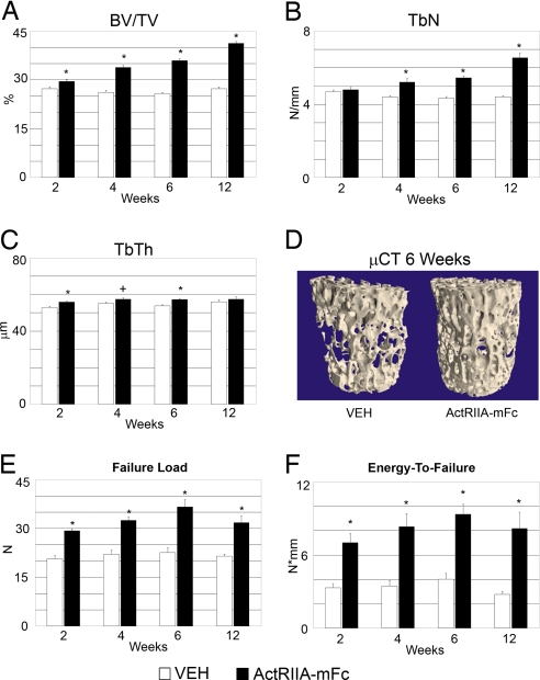
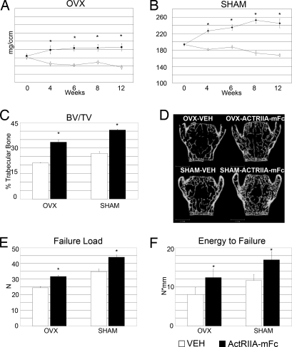
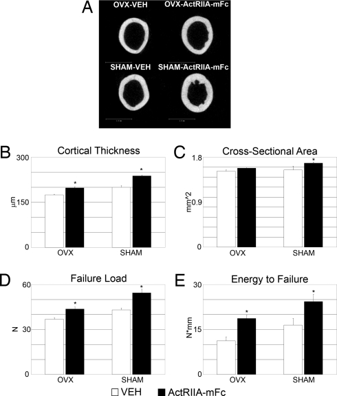
Diseases that affect the regulation of bone turnover can lead to skeletal fragility and increased fracture risk. Members of the TGF-beta superfamily have been shown to be involved in the regulation of bone mass. Activin A, a TGF-beta signaling ligand, is present at high levels in bone and may play a role in the regulation of bone metabolism. Here we demonstrate that pharmacological blockade of ligand signaling through the high affinity receptor for activin, type II activin receptor (ActRIIA), by administration of the soluble extracellular domain of ActRIIA fused to a murine IgG2a-Fc, increases bone formation, bone mass, and bone strength in normal mice and in ovariectomized mice with established bone loss. These observations support the development of this pharmacological strategy for the treatment of diseases with skeletal fragility.


Conflict of interest statement

Conflict of interest statement: R.S.P., M.C.-B., K.W.U., B.H., J.U., R.K., E.P., A.G., E.D.W., and J.S. are full-time employees of Acceleron Pharma.

Figures

Fig. 1.
Fig. 1.
ActRIIA–mFc treatment increases trabecular bone formation in normal mice. Static and dynamic histomorphometry parameters were measured in the distal femur. Open bars represent VEH-treated mice, and filled bars represent ActRIIA–mFc-treated mice. (A) Tb BV/TV (%). (B) Trabecular number (mm−1). (C) Trabecular thickness (μm). (D) ES/BS (%). (E) Nob/Bpm (mm−1). (F) Noc/Bpm (mm−1). (G) MS/BS (%). (H) Mineral apposition rate (μm/day). (I) Bone formation rate (μm3/μm2/day). (J) Representative images from von Kossa staining of the femurs at 6 weeks. (K and L) Serum osteocalcin levels (ng/ml) (K) and TRAP5b levels (units/liter) (L) as measured by ELISA. All results are presented as the mean ± SEM. *, P < 0.01.
Fig. 2.
Fig. 2.
Ex vivo μ and biomechanical analysis of the fifth lumbar vertebrae of ActRIIA–mFc-treated mice. Open bars represent VEH-treated mice, and filled bars represent ActRIIA–mFc-treated mice. (A) Tb BV/TV (%). (B) Trabecular number (mm−1). (C) TbTh (μm). (D) Representative three-dimensional μCT images of vertebrae from mice treated for 6 weeks with VEH or ActRIIA–mFc. (E) Vertebral failure load (N) from compression testing. (F) Energy-to-failure (N × nmm) from compression testing. All results are presented as the mean ± SEM. *, P < 0.01; +, P < 0.05.
Fig. 3.
Fig. 3.
ActRIIA–mFc treatment reverses trabecular bone loss in ovariectomized mice. OVX or SHAM mice were treated with ActRIIA–mFc (filled bars) or VEH (open bars) for a total of 12 weeks. In vivo pQCT assessment of trabecular bone density (mg/cm3) in the proximal tibia of OVX (A) and SHAM (B) mice was determined before dosing and after 4, 6, 8, and 12 weeks of treatment. (C) The Tb BV/TV (%) in OVX and SHAM mice was determined by ex vivo μCT analysis of the fifth lumbar vertebra. (D) Representative images of vertebrae are shown for each treatment group. (E) Vertebral failure load (N) determined by compression testing from OVX- and SHAM-treated mice. (F) Energy-to-failure (N × mm) from OVX and SHAM. All results are reported as the mean ± SEM. *, P < 0.01.
Fig. 4.
Fig. 4.
ActRIIA–mFc increases cortical bone volume and improves femoral strength. Cortical bone mass and strength was assessed in the femur of VEH-treated (open bars) and ActRIIA–mFc-treated (filled bars) mice by μCT after 12 weeks of treatment. (A) Representative cross-sectional images of the femoral diaphysis. (B) The cortical thickness (μm) at the diaphysis of OVX and SHAM mice. (C) Total cross-sectional area (mm2) of the femur in OVX and SHAM mice. (D) The failure load (N) was determined by three-point bending analysis of the femur for each group. (E) The energy-to-failure (N × mm) was also determined for each group. *, P ≤ 0.01.

References

    1. Rodan GA. Bone mass homeostasis and bisphosphonate action. Bone. 1997;20:1–4. - PubMed
    1. Lambrinoudaki I, Christodoulakos G, Botsis D. Bisphosphonates. Ann NY Acad Sci. 2006;1092:397–402. - PubMed
    1. Black DM, et al. The effects of parathyroid hormone and alendronate alone or in combination in postmenopausal osteoporosis. N Engl J Med. 2003;349:1207–1215. - PubMed
    1. MacDonald BR, Gowen M. Emerging therapies in osteoporosis. Best Pract Res Clin Rheumatol. 2001;15:483–496. - PubMed
    1. Burr D. Microdamage and bone strength. Osteoporosis Int. 2003;14(Suppl 5):67–72. - PubMed

MeSH terms

Substances