

AMPK alpha1 activation is required for stimulation of glucose uptake by twitch contraction, but not by H2O2, in mouse skeletal muscle
- PMID: 18461163
- PMCID: PMC2346549
- DOI: 10.1371/journal.pone.0002102
AMPK alpha1 activation is required for stimulation of glucose uptake by twitch contraction, but not by H2O2, in mouse skeletal muscle
Abstract
Background: AMPK is a promising pharmacological target in relation to metabolic disorders partly due to its non-insulin dependent glucose uptake promoting role in skeletal muscle. Of the 2 catalytic alpha-AMPK isoforms, alpha(2) AMPK is clearly required for stimulation of glucose transport into muscle by certain stimuli. In contrast, no clear function has yet been determined for alpha(1) AMPK in skeletal muscle, possibly due to alpha-AMPK isoform signaling redundancy. By applying low-intensity twitch-contraction and H(2)O(2) stimulation to activate alpha(1) AMPK, but not alpha(2) AMPK, in wildtype and alpha-AMPK transgenic mouse muscles, this study aimed to define conditions where alpha(1) AMPK is required to increase muscle glucose uptake.
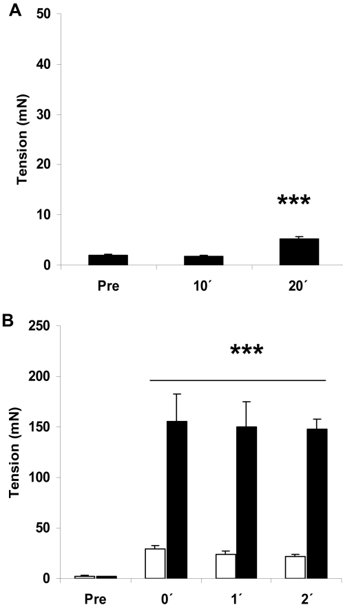
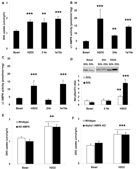
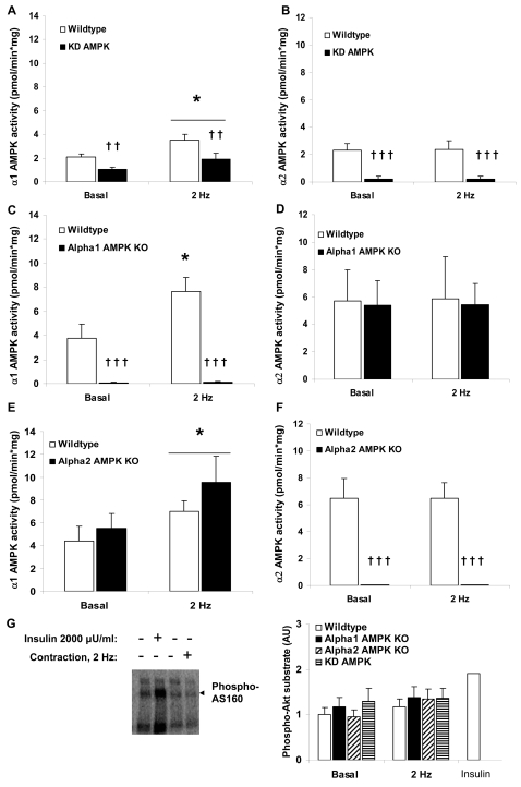
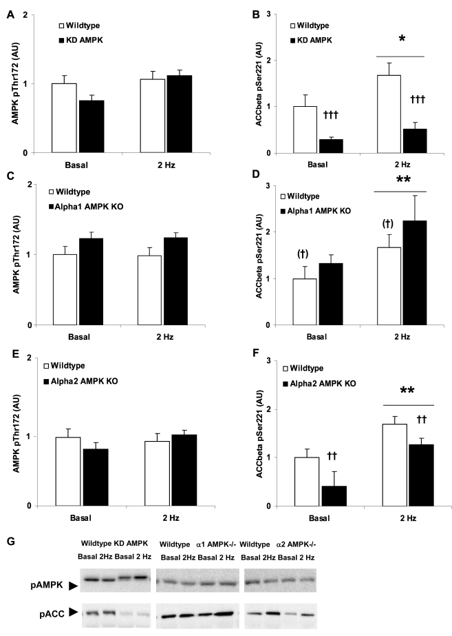
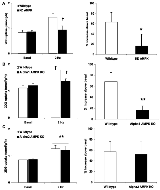
Methodology/principal findings: Following stimulation with H(2)O(2) (3 mM, 20 min) or twitch-contraction (0.1 ms pulse, 2 Hz, 2 min), signaling and 2-deoxyglucose uptake were measured in incubated soleus muscles from wildtype and muscle-specific kinase-dead AMPK (KD), alpha(1) AMPK knockout or alpha(2) AMPK knockout mice. H(2)O(2) increased the activity of both alpha(1) and alpha(2) AMPK in addition to Akt phosphorylation, and H(2)O(2)-stimulated glucose uptake was not reduced in any of the AMPK transgenic mouse models compared with wild type. In contrast, twitch-contraction increased the activity of alpha(1) AMPK, but not alpha(2) AMPK activity nor Akt or AS160 phosphorylation. Glucose uptake was markedly lower in alpha(1) AMPK knockout and KD AMPK muscles, but not in alpha(2) AMPK knockout muscles, following twitch stimulation.
Conclusions/significance: These results provide strong genetic evidence that alpha(1) AMPK, but not alpha(2) AMPK, Akt or AS160, is necessary for regulation of twitch-contraction stimulated glucose uptake. To our knowledge, this is the first report to show a major and essential role of alpha(1) AMPK in regulating a physiological endpoint in skeletal muscle. In contrast, AMPK is not essential for H(2)O(2)-stimulated muscle glucose uptake, as proposed by recent studies.
Conflict of interest statement
Figures
Similar articles
-
Knockout of the alpha2 but not alpha1 5'-AMP-activated protein kinase isoform abolishes 5-aminoimidazole-4-carboxamide-1-beta-4-ribofuranosidebut not contraction-induced glucose uptake in skeletal muscle.J Biol Chem. 2004 Jan 9;279(2):1070-9. doi: 10.1074/jbc.M306205200. Epub 2003 Oct 21. J Biol Chem. 2004. PMID: 14573616
-
Low-intensity contraction activates the alpha1-isoform of 5'-AMP-activated protein kinase in rat skeletal muscle.Am J Physiol Endocrinol Metab. 2006 Mar;290(3):E583-90. doi: 10.1152/ajpendo.00395.2005. Epub 2005 Oct 25. Am J Physiol Endocrinol Metab. 2006. PMID: 16249251
-
Possible CaMKK-dependent regulation of AMPK phosphorylation and glucose uptake at the onset of mild tetanic skeletal muscle contraction.Am J Physiol Endocrinol Metab. 2007 May;292(5):E1308-17. doi: 10.1152/ajpendo.00456.2006. Epub 2007 Jan 9. Am J Physiol Endocrinol Metab. 2007. PMID: 17213473
-
Physiological role of AMP-activated protein kinase (AMPK): insights from knockout mouse models.Biochem Soc Trans. 2003 Feb;31(Pt 1):216-9. doi: 10.1042/bst0310216. Biochem Soc Trans. 2003. PMID: 12546688 Review.
-
LKB1 and AMPK and the regulation of skeletal muscle metabolism.Curr Opin Clin Nutr Metab Care. 2008 May;11(3):227-32. doi: 10.1097/MCO.0b013e3282fb7b76. Curr Opin Clin Nutr Metab Care. 2008. PMID: 18403917 Free PMC article. Review.
Cited by
-
The physiological regulation of glucose flux into muscle in vivo.J Exp Biol. 2011 Jan 15;214(Pt 2):254-62. doi: 10.1242/jeb.048041. J Exp Biol. 2011. PMID: 21177945 Free PMC article.
-
LKB1 regulates lipid oxidation during exercise independently of AMPK.Diabetes. 2013 May;62(5):1490-9. doi: 10.2337/db12-1160. Epub 2013 Jan 24. Diabetes. 2013. PMID: 23349504 Free PMC article.
-
Insulin down-regulates the expression of ubiquitin E3 ligases partially by inhibiting the activity and expression of AMP-activated protein kinase in L6 myotubes.Biosci Rep. 2015 Jul 20;35(4):e00242. doi: 10.1042/BSR20150017. Biosci Rep. 2015. PMID: 26193886 Free PMC article.
-
Energy (and Reactive Oxygen Species Generation) Saving Distribution of Mitochondria for the Activation of ATP Production in Skeletal Muscle.Antioxidants (Basel). 2023 Aug 17;12(8):1624. doi: 10.3390/antiox12081624. Antioxidants (Basel). 2023. PMID: 37627619 Free PMC article. Review.
-
β-Actin shows limited mobility and is required only for supraphysiological insulin-stimulated glucose transport in young adult soleus muscle.Am J Physiol Endocrinol Metab. 2018 Jul 1;315(1):E110-E125. doi: 10.1152/ajpendo.00392.2017. Epub 2018 Mar 13. Am J Physiol Endocrinol Metab. 2018. PMID: 29533739 Free PMC article.
References
-
- Kahn BB, Alquier T, Carling D, Hardie DG. AMP-activated protein kinase: Ancient energy gauge provides clues to modern understanding of metabolism. Cell Metabolism. 2005;1:15–25. - PubMed
-
- Fujii N, Hirshman MF, Kane EM, Ho RC, Peter LE, et al. AMP-activated Protein Kinase {alpha}2 Activity Is Not Essential for Contraction- and Hyperosmolarity-induced Glucose Transport in Skeletal Muscle. J Biol Chem. 2005;280:39033–39041. - PubMed
-
- Jørgensen SB, Viollet B, Andreelli F, Frosig C, Birk JB, et al. Knockout of the alpha2 but Not alpha1 5′-AMP-activated Protein Kinase Isoform Abolishes 5-Aminoimidazole-4-carboxamide-1-beta-4-ribofuranosidebut Not Contraction-induced Glucose Uptake in Skeletal Muscle. J Biol Chem. 2004;279:1070–1079. - PubMed
-
- Mu J, Brozinick JT, Jr., Valladares O, Bucan M, Birnbaum MJ. A role for AMP-activated protein kinase in contraction- and hypoxia-regulated glucose transport in skeletal muscle. Mol Cell. 2001;7:1085–1094. - PubMed
-
- Kramer HF, Witczak CA, Fujii N, Jessen N, Taylor EB, et al. Distinct Signals Regulate AS160 Phosphorylation in Response to Insulin, AICAR, and Contraction in Mouse Skeletal Muscle. Diabetes. 2006;55:2067–2076. - PubMed
Publication types
MeSH terms
Substances
LinkOut - more resources
Full Text Sources
Molecular Biology Databases
