. 2008 May 7;3(5):e2122.
doi: 10.1371/journal.pone.0002122.

Differentiation of human embryonic stem cells to regional specific neural precursors in chemically defined medium conditions

Affiliations

Differentiation of human embryonic stem cells to regional specific neural precursors in chemically defined medium conditions

Slaven Erceg et al. PLoS One. .

Abstract

Background: Human embryonic stem cells (hESC) provide a unique model to study early events in human development. The hESC-derived cells can potentially be used to replace or restore different tissues including neuronal that have been damaged by disease or injury.

Methodology and principal findings: The cells of two different hESC lines were converted to neural rosettes using adherent and chemically defined conditions. The progenitor cells were exposed to retinoic acid (RA) or to human recombinant basic fibroblast growth factor (bFGF) in the late phase of the rosette formation. Exposing the progenitor cells to RA suppressed differentiation to rostral forebrain dopamine neural lineage and promoted that of spinal neural tissue including motor neurons. The functional characteristics of these differentiated neuronal precursors under both, rostral (bFGF) and caudalizing (RA) signals were confirmed by patch clamp analysis.

Conclusions/significance: These findings suggest that our differentiation protocol has the capacity to generate region-specific and electrophysiologically active neurons under in vitro conditions without embryoid body formation, co-culture with stromal cells and without presence of cells of mesodermal or endodermal lineages.


Conflict of interest statement

Competing Interests: The authors have declared that no competing interests exist.

Figures

Figure 1
Figure 1. Differentiation of hESC to neural progenitors.
(A) Schematic representation of the different steps in feeder-free and chemically defined medium conditions (see Material and Methods). (B and C) After two days in adherent animal-free substrate and modified TESR1 medium the cells were organized into rosettes (indicated with black arrows, also inserts, B and C). (D–F) RT-PCR analysis indicates changes in gene expression of pluripotency and neural markers through three steps of differentiation protocols: (D) oligodendrocyte and astrocytes expression profile; (E) Changes in gene expression of main pluripotency markers and general neural markers. (F) Semiquantitative RT-PCR of some endodermal, mesodermal, epidermal, and trophoblast markers. Bars: (B) 100 µm; (C) 200 µm.
Figure 2
Figure 2. Immunocytochemical characterizations of hESC-derived neuronal precursors at D28.
The cells are positive for neural progenitor markers, such as (A) Tuj1 (green), (B) Musashi (red), (C) A2B5 (green), (D) MAP2 (red coexpressed with Tuj1). Blue indicates DAPI. Bars: (A–C) 50 µm; (D) 25 µm.
Figure 3
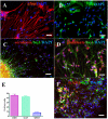
Figure 3. hESC derived neural progenitors give rise to (A) astrocytes (GFAP red) and (B) oligodendrocytes (O4, green).
The cells are serotonin+ (C; red coexpressed with Tuj1), glutamate+ (D; red, coexpressed with Tuj1), and GABA+ (F; red, coexpressed with Tuj1). Blue indicates DAPI. GFAP+ and O4+ cells are stained at D56. Bars: (A, B, D) 25 µm; (C, F) 50 µm. (E) Data were averaged and represented as means±S.E.M.
Figure 4
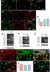
Figure 4. Neuronal specification of cells obtained using protocols with or without RA.
The hESC derived neural progenitors display rostral phenotype: (A) the cells are Gbx2+ (red, coexpressed with Tuj1) and (B) OTX2+ (red, coexpressed with Tuj1). (C) Almost all GABA+ cells (red) are TH+ (green). (D) The percentage of the cells positive for rostral markers. (E) RT-PCR analysis of rostral markers of hESC-derived neural progenitors with or without RA. (F and G) RT-PCR analysis of spinal cord markers. The hESC derived neural progenitors treated with RA display spinal cord phenotype; (H) almost all HB9 cells (red) are cholinergic (green). (I) The cells are mostly HB9+ (red) and Tuj1+ (green) positive. (J) Percentage of HB9+, Chat+, and HOXC8+ cells. (K) The cells are also HOXC8+ (green). Blue indicates DAPI. Bars: (A) 50 µm; (B, C, H, I, K) 100 µm. (D, J) Data were averaged and represented as means±S.E.M.
Figure 5
Figure 5. Neuronal excitability and study of voltage-dependent channels in the newly generated neurons by bFGF and RA treatment.
(A) Action potentials evoked by depolarizing current steps (40 pA) in two different neurons resulted in different firing patterns. In both cases, the spikes were fully blocked with 1 µM TTX (red). (B) Family of voltage-dependent currents obtained from a –30 mV to +50 mV in 10 mV increasing steps protocol from a Vh = −70 mV. An early inward current suggests the presence of voltage-dependent sodium channels, while the second outward component is consistent with the presence of voltage-dependent K+ channels, as seen in the I-V relation. (C) Blockade of TTX-sensitive voltage-dependent Na+ channel. The early component present in (B) was fully blocked with 1 µM TTX (red trace) (n = 39). (D) The outward current was blocked by 2 mM 4-AP (red trace) (n = 10).
Figure 6
Figure 6. Neurotransmitter sensitivity.
(A) Response to 100 µM glutamate (Glu) and blockade of the current by the simultaneous application of the antagonist CNQX (n = 11). The horizontal bar indicates the duration of agonist (red line) and/or antagonist (blue line) or modulator (green line) application. (B): Response of cells to 100 µM of GABA and blocking of the current with the specific GABAA receptor antagonist bicuculline (Bic) (n = 25). (C): Reversible downregulation of Glu currents in cells treated with bFGF by applying 1 mM of the metabotropic neurotransmitter dopamine (DA) for two minutes to the cells (n = 8). (D): Response of cells treated with RA to 100 µM acetylcholine (Ach) (n = 8). The current was fully blocked with 100 µM of (−) tubocurarine (Cur) (n = 6).

References

    1. Odorico JS, Kaufman DS, Thomson JA. Multilineage differentiation from human embryonic stem cell lines. Stem Cells. 2001;19:193–204. - PubMed
    1. Schuldiner M, Eiges R, Eden A, Yanuka O, Itskovitz-Eldor J, et al. Induced neuronal differentiation of human embryonic stem cells. Brain Res. 2001;913:201–205. - PubMed
    1. Zhang SC, Wernig M, Duncan ID, Brustle O, Thomson JA. In vitro differentiation of transplantable neural precursors from human embryonic stem cells. Nat Biotechnol. 2001;19:1129–1133. - PubMed
    1. Keirstead HS, Nistor G, Bernal G, Totoiu M, Cloutier F, et al. Human embryonic stem cell-derived oligodendrocyte progenitor cell transplants remyelinate and restore locomotion after spinal cord injury. J Neurosci. 2005;25:4694–4705. - PMC - PubMed
    1. Carpenter MK, Inokuma MS, Denham J, Mujtaba T, Chiu CP, et al. Enrichment of neurons and neural precursors from human embryonic stem cells. Exp Neurol. 2001;172:383–397. - PubMed

Publication types