. 2008 May 7;3(5):e2115.
doi: 10.1371/journal.pone.0002115.

Myosin VIIA, important for human auditory function, is necessary for Drosophila auditory organ development

Affiliations

Myosin VIIA, important for human auditory function, is necessary for Drosophila auditory organ development

Sokol V Todi et al. PLoS One. .

Abstract

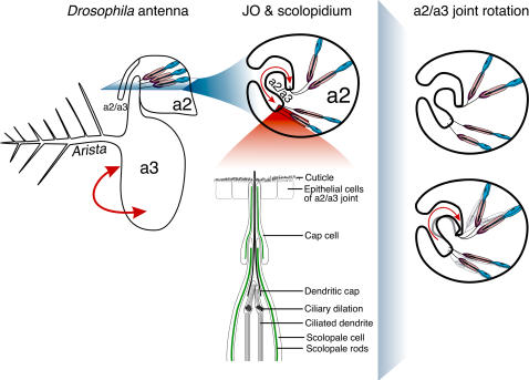
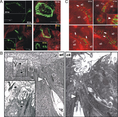
Background: Myosin VIIA (MyoVIIA) is an unconventional myosin necessary for vertebrate audition [1]-[5]. Human auditory transduction occurs in sensory hair cells with a staircase-like arrangement of apical protrusions called stereocilia. In these hair cells, MyoVIIA maintains stereocilia organization [6]. Severe mutations in the Drosophila MyoVIIA orthologue, crinkled (ck), are semi-lethal [7] and lead to deafness by disrupting antennal auditory organ (Johnston's Organ, JO) organization [8]. ck/MyoVIIA mutations result in apical detachment of auditory transduction units (scolopidia) from the cuticle that transmits antennal vibrations as mechanical stimuli to JO.

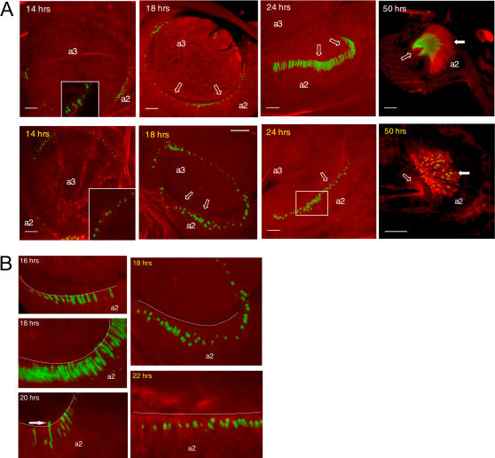
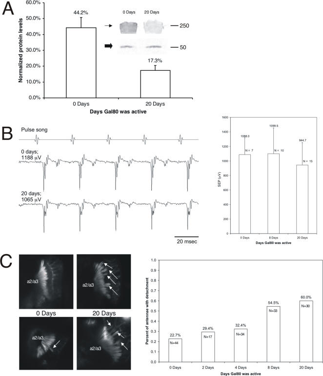
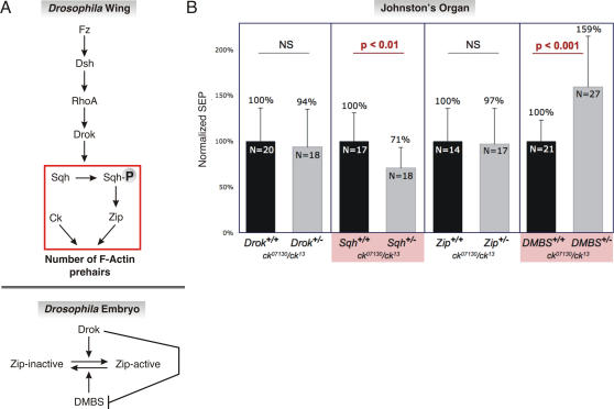
Principal findings: Using flies expressing GFP-tagged NompA, a protein required for auditory organ organization in Drosophila, we examined the role of ck/MyoVIIA in JO development and maintenance through confocal microscopy and extracellular electrophysiology. Here we show that ck/MyoVIIA is necessary early in the developing antenna for initial apical attachment of the scolopidia to the articulating joint. ck/MyoVIIA is also necessary to maintain scolopidial attachment throughout adulthood. Moreover, in the adult JO, ck/MyoVIIA genetically interacts with the non-muscle myosin II (through its regulatory light chain protein and the myosin binding subunit of myosin II phosphatase). Such genetic interactions have not previously been observed in scolopidia. These factors are therefore candidates for modulating MyoVIIA activity in vertebrates.

Conclusions: Our findings indicate that MyoVIIA plays evolutionarily conserved roles in auditory organ development and maintenance in invertebrates and vertebrates, enhancing our understanding of auditory organ development and function, as well as providing significant clues for future research.


Conflict of interest statement

Competing Interests: The authors have declared that no competing interests exist.

Figures

Figure 1
Figure 1. Schematic of the antenna and JO.
Drawing not to scale.
Figure 2
Figure 2. ck/MyoVIIA is necessary for JO organization during development.
A: Comparison of control (ck13/CyO; white labels) vs. mutant (ck13/ck13; yellow labels) developing JO. Green channel: GFP-NompA labelling dendritic caps. Red channel: Texas red-phalloidin labelling actin filaments. Block arrows: developing scolopale rods. Open block arrows: direction of a2/a3 boundary. Box indicates disorganization of cap alignment in JO. Inset in 14 hrs control: in some cases the cap shows an elongated profile by this time. Inset in 14 hrs mutant antenna: magnification of the globular profile of the cap. Scale bar = 20 µm. B: Similar staining and labelling as “A”. Note dendritic caps juxtaposed with the perimeter of a2 (dotted line) or extending into the space between a2/a3 in controls (arrow; white labels). In mutants (yellow labels) the caps remain distanced from the future a2/a3 joint.
Figure 3
Figure 3. Scolopidia lacking ck/MyoVIIA are not attached to the a2/a3 joint during development.
A: Wild type (ck13/CyO; white labels) and mutant (ck13/ck13; yellow labels) cultured antennal discs labelled as in Figure 2 support our finding that ck/MyoVIIA is necessary for JO organization. B: Representative EM micrographs from wild type (26 hrs APF) and mutant (28 hrs APF) antennae. Black arrowheads: dendritic caps extending beyond the a2/a3 joint boundary in controls. White arrowheads: dendritic caps at the apical levels of the scolopale space. In ck mutants the cap is more compact than in controls. Inset: magnification of the boxed area. C: Wild type (ck13/CyO; white labels) and mutant (ck13/ck13; yellow labels) cultured antennal discs labelled with anti-cadherin antibody. Green channel: GFP-NompA. Red channel: anti-cadherin. Arrows: cellular junction “tracts” in mutant discs. Open block arrows: a2/a3 boundary. Inset: enlargement of the boxed area. Arrowhead: a dendritic cap extending beyond the a2/a3 boundary. Block arrows: cell-cell junctions.
Figure 4
Figure 4. ck/MyoVIIA is important for maintaining adult JO organization.
A: Histogram of densitometry analysis of the western blot (inset) staining for ck/MyoVIIA (arrow) and β-tubulin (block arrow; loading control) from flies with active Gal80 for 0 or 20 days. Mean +/− SD; N = 4. B: Representative electrophysiological recordings (left) from the antennal nerve of flies with active Gal80 for 0, 8 or 20 days show no significant difference. Right: histograms of recordings. Mean +/− SD. C: JO organization in adults. Arrows indicate detachment. Histograms show percent of antennae with detached scolopidia.
Figure 5
Figure 5. ck/MyoVIIA function is modulated by Sqh and DMBS in adult JO.
A: Genetic interactions of ck/MyoVIIA with actin pathways in the Drosophila wing (top panel) and in embryonic dorsal closure (bottom panel). Boxed area indicates the portion of the pathway investigated in panel B. Based on , . B: ck/MyoVIIA genetically interacts with Sqh and DMBS in adult JO. Responses from flies with only one functional copy of Drok, sqh, zip/MyoII or DMBS were normalized to their respective, two-copy sibling controls, all in a sensitized ck/MyoVIIA background. Histograms show mean +/− SD.

References

    1. Ernest S, Rauch G-J, Haffter P, Geisler R, Petit C, et al. Mariner is defective in myosin VIIA: a zebrafish model for human hereditary deafness. Hum Mol Genet. 2000;9:2189–2196. - PubMed
    1. Gibson F, Walsh J, Mburu P, Varela A, Brown KA, et al. A type VII myosin encoded by the mouse deafness gene shaker-1. Nature. 1995;374:62–64. - PubMed
    1. Liu X-Z, Walsh J, Mburu P, Kendrick-Jones J, Cope MJTV, et al. Mutations in the myosin VIIA gene cause non-syndromic recessive deafness. Nature Genet. 1997;16:188–190. - PubMed
    1. Liu X-Z, Walsh J, Tamagawa Y, Kitamura K, Nishizawa M, et al. Autosomal dominant non-syndromic deafness caused by a mutation in the myosin VIIA gene. Nature Genet. 1997;17:268–269. - PubMed
    1. Weil D, Blanchard S, Kaplan J, Guilford P, Gibson F, et al. Defective myosin VIIA gene responsible for Usher syndrome type 1B. Nature. 1995;374:60–61. - PubMed

Publication types