Review
. 2009 Jan;58(1-2):105-34.
doi: 10.1007/s00285-008-0182-2. Epub 2008 May 7.

Mathematics of cell motility: have we got its number?

Affiliations
Review

Mathematics of cell motility: have we got its number?

Alex Mogilner. J Math Biol. 2009 Jan.

Abstract

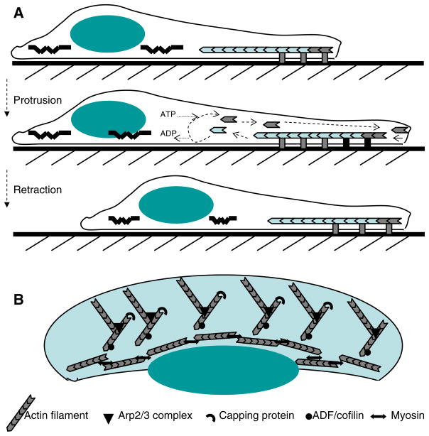
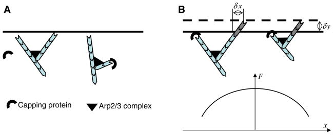
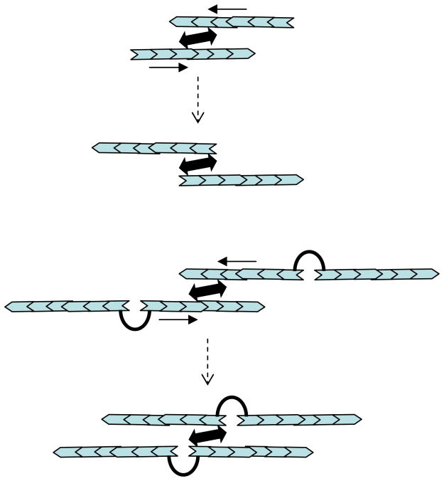
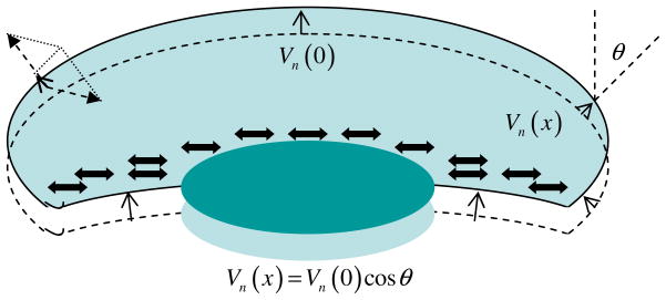
Mathematical and computational modeling is rapidly becoming an essential research technique complementing traditional experimental biological methods. However, lack of standard modeling methods, difficulties of translating biological phenomena into mathematical language, and differences in biological and mathematical mentalities continue to hinder the scientific progress. Here we focus on one area-cell motility-characterized by an unusually high modeling activity, largely due to a vast amount of quantitative, biophysical data, 'modular' character of motility, and pioneering vision of the area's experimental leaders. In this review, after brief introduction to biology of cell movements, we discuss quantitative models of actin dynamics, protrusion, adhesion, contraction, and cell shape and movement that made an impact on the process of biological discovery. We also comment on modeling approaches and open questions.


Figures

Fig. 1
Fig. 1
Schematic cell motility cycle; a side view, b top view
Fig. 2
Fig. 2
Polymerization ratchet model: rapid thermal undulations of both filament and membrane are ratcheted by the intercalation of actin monomers into the gap between the barbed end and membrane. Notations are explained in the text
Fig. 3
Fig. 3
a Angular organization of the lamellipodial actin array: top ‘Mother and daughter’ filaments grow at 70° relative to each other because of Arp2/3 geometry. Barbed ends growing at approximately 35° relative to the protrusion direction are close to the cell membrane and protected from capping. If the mother filament is almost normal to the membrane, then the daughter filament is almost normal to the protrusion direction, lags behind, is capped rapidly, and does not branch out next generation filaments. These processes cause angular symmetry of the actin array. b Spatial (lateral) organization of the lamellipodial actin array. Top advancement of the leading edge (δy) causes the lateral flow of the growing barbed ends (δx). Together with two other processes (capping and Arp2/3-mediated branching), these lead to the inverted parabolic distribution of the F-actin density along the leading edge (bottom)
Fig. 4
Fig. 4
Funneling effect: a relatively small number of the uncapped filaments grow mostly at the leading edge. There are many more disassembling pointed ends across the lamellipod. The monomer flux from many pointed ends is funneled to the relatively few barbed ends
Fig. 5
Fig. 5
Microscopic actin-myosin contraction. Top motor heads of bi-polar myosin cluster try to glide toward respective barbed end, and as a result pull the filaments in the direction of their pointed ends until the barbed ends are co-localized. In the end, the radius of gyration of the filament pair is usually greater than it was in the beginning, so contraction is not achieved. Bottom if the barbed ends are crosslinked by a binding protein, then two interacting filament pairs glide until all barbed ends are co-localized, so myosin contracts actin
Fig. 6
Fig. 6
Adhesion dynamics: adhesions assemble rapidly and hierarchically at the leading edge to ensure firm adhesion at the front, and then slowly ‘age’—disassemble—so toward the rear the adhesion strength diminishes. Only few essential adhesion molecules are shown; the order and rate of assembly/disassembly are not to be taken literally
Fig. 7
Fig. 7
‘Spring-and-dashpot’ 1D motility model
Fig. 8
Fig. 8
Graded radial extension model and actin growth/myosin contraction mechanism. Sum of polymerization and centripetal actin flow (dashed arrows) result in the vector, projection of which at the direction locally normal to the cell boundary (illustrated at the left front) gives the resulting local extension rate, Vn (x). The steady shape of the leading edge is defined by the relation Vn (x) = Vn (0) cos θ(x). Similar relations are applicable at the rear edge. In the framework of the moving cell, myosin is swept to the rear, contracts visco-elastic actin network and generates centripetal actin flow that is weak at the front and strong at the sides and rear. This allows myosin to pull up the rear, restrain the sides and let the leading edge protrude
Fig. 9
Fig. 9
Fragment motility. Lamellipodial fragment excised from the whole cell assumes stationary discoid shape. Weak transient mechanical perturbation by fluid flow does not break the fragment symmetry. Strong persistent perturbation causes symmetry break and consequent locomotion of crescent-like fragment
Fig. 10
Fig. 10
Listeria motility. Radial growth and resulting deformations of the actin comet-like tail generate elastic ‘squeezing’ stress propelling the cell forward. Inset dendritic actin array at the rear surface of the cell

Similar articles

Cited by

References

    1. Abercrombie M, Heaysman JE, Pegrum SM. The locomotion of fibroblasts in culture. I. Movements of the leading edge. Exp Cell Res. 1970;59:393–398. - PubMed
    1. Abraham VC, Krishnamurthi V, Taylor DL, Lanni F. The actin-based nanomachine at the leading edge of migrating cells. Biophys J. 1999;77:1721–1732. - PMC - PubMed
    1. Alberts JB, Odell GM. In silico reconstitution of Listeria propulsion exhibits nano-saltation. PLoS Biol. 2004;2:e412. - PMC - PubMed
    1. Alt W, Dembo M. Cytoplasm dynamics and cell motion: two-phase flow models. Math Biosci. 1999;156:207–228. - PubMed
    1. Asthagiri AR, Lauffenburger DA. Bioengineering models of cell signaling. Annu Rev Biomed Eng. 2000;2:31–53. - PubMed

Publication types

LinkOut - more resources