. 2008 Jun;38(6):1533-47.
doi: 10.1002/eji.200737909.

IL-10-dependent partial refractoriness to Toll-like receptor stimulation modulates gut mucosal dendritic cell function

Affiliations
Free PMC article

IL-10-dependent partial refractoriness to Toll-like receptor stimulation modulates gut mucosal dendritic cell function

Ivan Monteleone et al. Eur J Immunol. 2008 Jun.
Free PMC article

Abstract

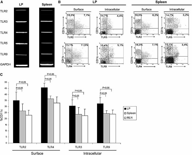
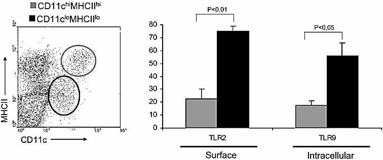
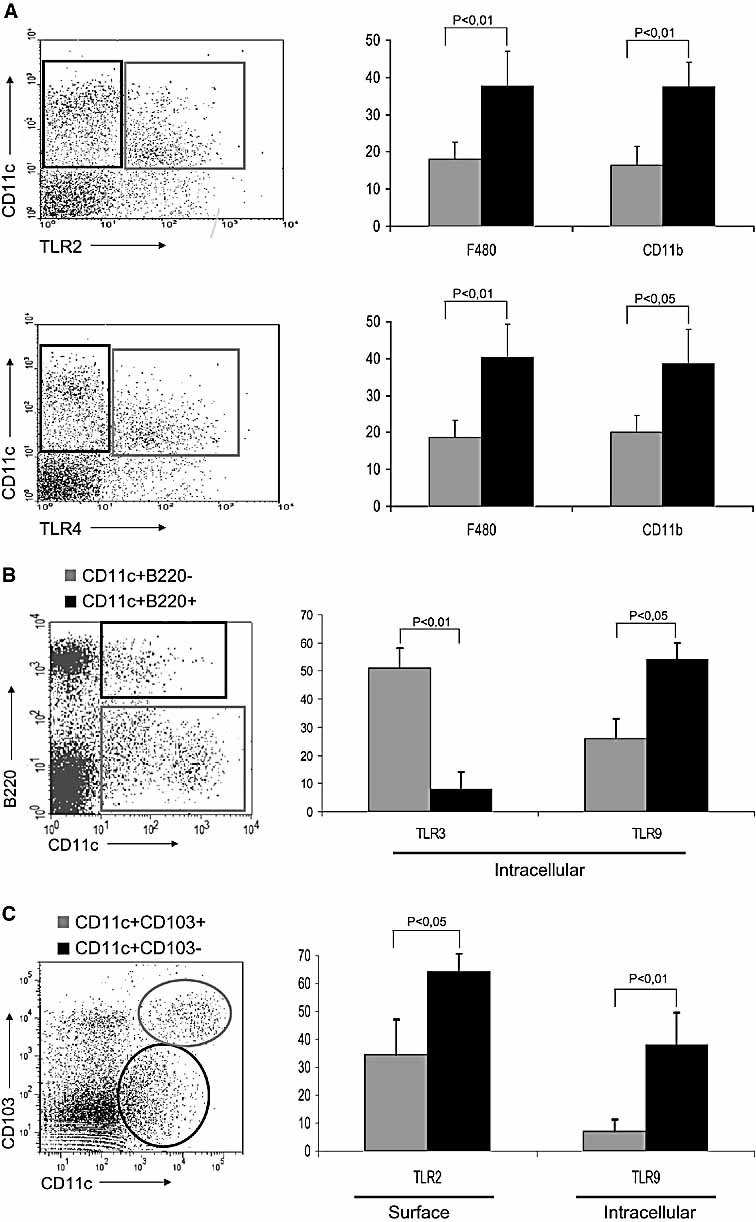
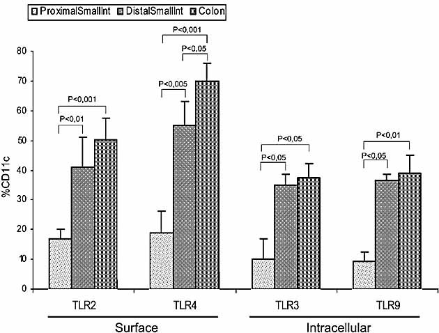
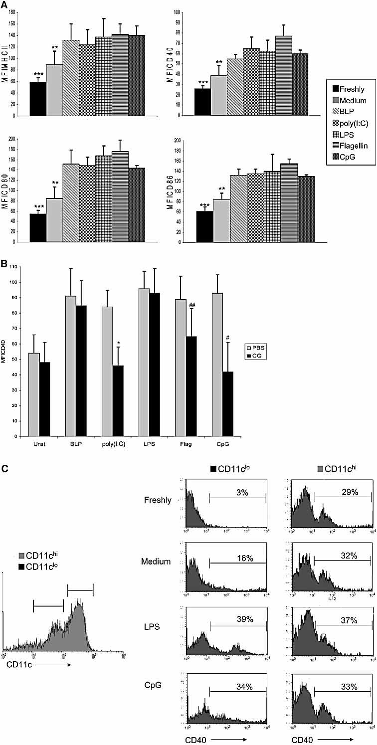
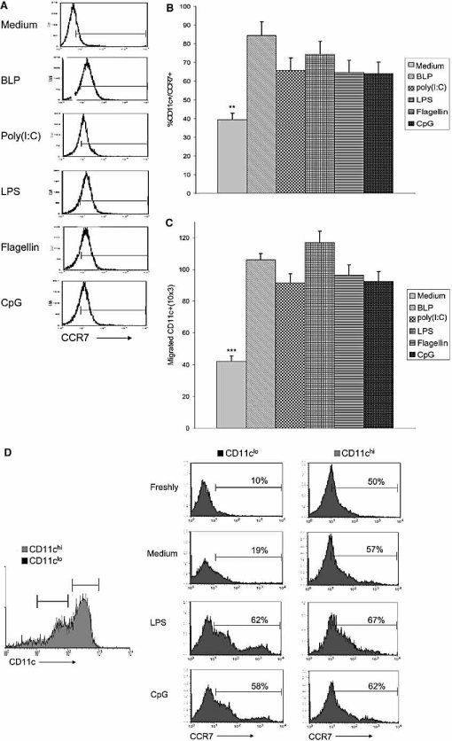
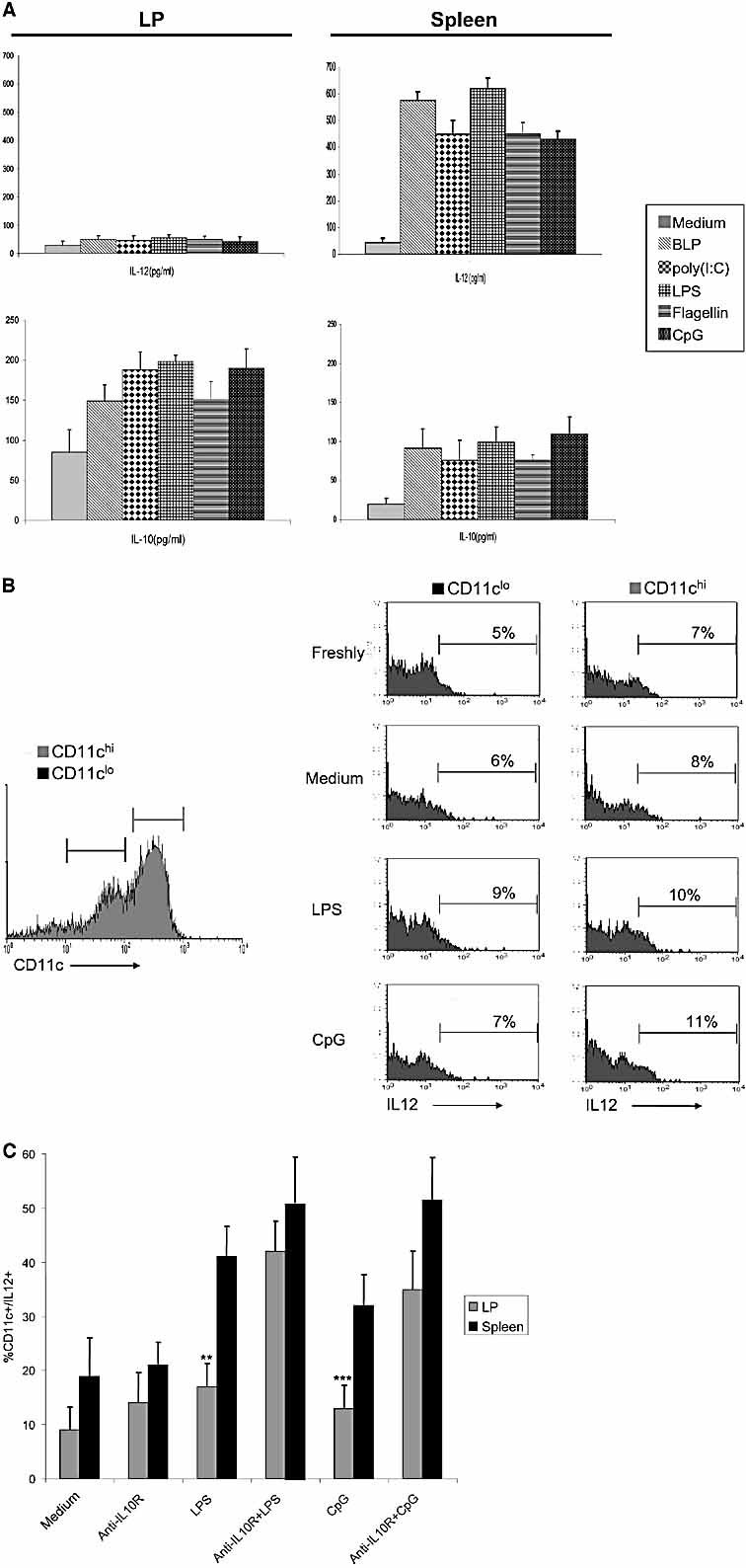
The default response of the intestinal immune system to most antigens is the induction of immunological tolerance, which is difficult to reconcile with the constant exposure to ligands for TLR and other pattern recognition receptors. We showed previously that dendritic cells (DC) from the lamina propria of normal mouse intestine may be inherently tolerogenic and here we have explored how this might relate to the expression and function of Toll-like receptors (TLR). Lamina propria (LP) DC showed higher levels of TLR 2, 3, 4 and 9 protein expression than spleen and MLN DC, with most TLR-expressing DC in the gut being CD11c(lo), class II MHC(lo), CD103(-), CD11b(-) and F4/80(-). TLR expression by lamina propria DC was low in the upper small intestine and higher in distal small intestine and colon. Freshly isolated lamina propria DC expressed some CD40, CD80, CD86 and functional CCR7. These were up-regulated on CD11c(lo), but not on CD11c(hi) LP DC by stimulation via TLR. However, there was little induction of IL-12 by either subset in response to TLR ligation. This was associated with constitutive IL-10 production and was reversed by blocking IL-10 function. Thus, IL-10 may maintain LP DC in a partially unresponsive state to TLR ligation, allowing them to have a critical role in immune homeostasis in the gut.


Figures

Figure 1
Figure 1
TLR expression by DC in LP, spleen and MLN. (A) mRNA for TLR2, 3, 4, 5, 9 and GAPDH was identified by RT-PCR using MACS-purified CD11c+ cells isolated by enzymatic digestion from the small intestinal LP and spleen. The results are representative of two repeat experiments. (B) Representative flow cytometric analysis showing the surface expression of TLR2 and 4 and the intracellular expression of TLR3 and 9 by small intestinal LP and spleen DC. Cells were isolated by enzymatic digestion, stained with FITC or PE antibodies anti-CD11c and FITC or PE antibodies anti-TLR, with dead cells gated out by staining with PI. The data shown are the proportion of live gated, CD11c+ cells positive for each marker. (C) Proportions of DC expressing surface TLR2 or 4, and intracellular TLR3 or 9 in LP (black), spleen (grey) and MLN (white) as assessed by flow cytometric analysis as described in (B). The data shown are the percentages of TLR+ cells out of live-gated (PI) CD11c+ cells in each tissue and are the mean + 1 SD of three separate experiments.
Figure 2
Figure 2
Two major sub-populations of CD11clo class II MHClo and CD11chi class II MHChi DC were identified among freshly isolated, enzymatically digested small intestinal LP cells (left panel). The proportions of live gated CD11clo class II MHClo (black) and CD11chi class II MHChi (grey) DC expressing surface TLR2 or intracellular TLR 9 are shown (right panel). The data shown are the percentages of TLR+ cells out of live-gated (PI) CD11c+ cells in each tissue and are the mean ± 1 SD of three separate experiments.
Figure 3
Figure 3
Distribution of TLR expression in different LP DC subsets. (A) TLR are expressed mainly by LP DC (CD11c+CD11bF4/80). CD11c+TLR2 and CD11c+TLR2+ subsets of freshly-isolated LP cells were shown in the top left panel. The proportions of live-gated CD11c+TLR2 (black) and CD11c+TLR2+ (grey) DC expressing surface F4/80 or CD11b are shown in the top right panel. CD11c+TLR4 and CD11c+TLR4+ subsets of freshly-isolated LP cells were shown in the bottom left panel. The proportions of live-gated CD11c+TLR4 (black) and CD11c+TLR4+ (grey) DC expressing surface F4/80 or CD11b are shown in the bottom right panel. (B) Expression of TLR by the CD11c+B220 and CD11c+B220+ DC subsets within the small intestinal LP cells (left panel). The proportions of live-gated CD11c+B220+ (black) and CD11c+B220 (grey) DC expressing intracellular TLR3 or TLR9 are shown (right panel). (C) Two subpopulation of DC CD11c+CD103+ (grey) and CD11c+ CD103 (black) were found in freshly isolated small intestinal LP cells (left panel). The percentages of gated CD11c+CD103+ (grey) and CD11c+CD103 (black) DC expressing intracellular TLR9 or surface TLR3 are shown (right panel). The data shown are the percentages of F4/80+, CD11b+ or TLR+ cells out of live-gated (PI) CD11c+ cells in each tissue and are the mean + 1 SD of three separate experiments.
Figure 4
Figure 4
TLR expression on LP DC at different anatomical locations in the intestine. The proportions of freshly isolated, enzymatically digested small intestinal CD11c+ LP cells expressing surface TLR2 or 4, and intracellular TLR 3 or 9 in the proximal or distal small intestine or colon. All data are the mean + 1 SD of three separate experiments.
Figure 5
Figure 5
Effects of TLR ligation on the expression of activation markers by LP DC. (A) MACS-purified CD11c+ cells isolated by enzymatic digestion from the small intestinal LP were cultured with medium alone, or with different TLR ligands. After 12 h, DC were analysed for the expression of CD40, CD80, CD86 and class II MHC by flow cytometry. Freshly isolated DC were analysed in parallel as controls. The graph shows the MFI for each marker on live gated CD11c+ cells (***p≤0.05 vs. medium-treated and p<0.01 vs. all TLR ligands; **p≤0.05 vs. all TLR ligands). (B) Activation of DC by intracellular TLR requires endosomal acidification. MACS-purified CD11c+ LP DC were pre-treated with 2.5 μg/mL chloroquine (CQ, black) or PBS (grey) for 15 min and then stimulated for 12 h with different TLR ligands, prior to CD40 expression analysis by flow cytometry. The graph shows the MFI of CD40 expression on live gated CD11c+ cells (*p<0.005 vs. CD11c+ cells pretreated with PBS and stimulated with poly(I:C); #p<0.01 vs. CD11c+ cells pre-treated with PBS and stimulated with CpG; ##p<0.05 vs. CD11c+ cells pretreated with PBS and stimulated with flagellin). The data are the mean + 1 SD of three separate experiments. (C) CD11clo and CD11chi LP DC differ in their response to TLR ligands. LP cells were FACS-sorted into CD11chi (grey line) and CD11clo (black line) subsets and the histograms show CD40 expression on freshly isolated cells, or after incubation in medium alone, LPS or CpG for 12 h.
Figure 6
Figure 6
TLR stimulation enhances functional CCR7 expression by LP DC. (A) Representative flow cytometric analysis of CCR7 expression on live gated, MACS-purified CD11c+ cells from small intestinal LP stained with PE anti-CCR7 after stimulation for 6 h with medium alone, or with different TLR ligands. (B) Overall levels of expression of CCR7 on live gated, MACS-purified CD11c+ cells from small intestinal LP after stimulation for 6 h with medium alone or with different TLR ligands (**p<0.001 vs. BLP and LPS stimulation, p<0.005 vs. all other TLR stimulations). The data are the means + 1 SD of three separate experiments. (C) Chemotaxis of LP DC to CCR7 ligand CCL19. MACS-purified CD11c+ cells from small intestinal LP were cultured in medium, or with different TLR ligands for 4 h before being placed on the top of a transwell membrane and allowed to migrate in response to CCL19 (MIP-3β) in the bottom chamber. The number of CD11c+ cells which had migrated into the bottom chamber was determined by flow cytometry (***p≤0.001 vs. all TLR ligands). (D) CD11clo and CD11chi LP DC differ in their response to TLR ligands. LP cells were FACS-sorted into CD11chi (grey line) and CD11clo (black line) subsets and the histograms show CCR7 expression on freshly isolated cells, or after incubation in medium alone, LPS or CpG for 12 h.
Figure 7
Figure 7
Constitutive production of IL-10 inhibits inflammatory cytokine production by LP DC in response to TLR stimulation. (A) Production of IL-12p70 (top) and IL-10 (bottom) by purified MACS-purified CD11c+ DC from small intestinal LP (left) or spleen (right) after culture with medium alone or with different TLR ligands for 24 h. Supernatants were analysed by sandwich ELISA and the results shown are pg/mL for duplicate cultures and are the means + 1 SD of three separate experiments. (B) Both CD11clo and CD11chi LP DC fail to produce IL-12 in response to TLR ligands. LP cells were FACS-sorted into CD11chi (grey line) and CD11clo (black line) subsets and the histograms show intracellular expression of IL-12p40 by freshly isolated cells, or after overnight incubation with medium alone, LPS or CpG for 6 h. (C) MACS-purified CD11c+ DC from small intestinal LP or spleen were pre-treated with medium alone or with 2 ng/mL of anti-IL10 receptor antibody for 60 min before being stimulated with TLR ligands for 6 h. Intracellular IL-12p70 levels were analysed by flow cytometry and the data shown are the percentage live gated CD11c+ cells positive for IL-12 and are the mean + 1 SD of three separate experiments. **p≤0.01 vs. LP DC preincubated with anti-IL10R and LPS; ***p<0.05 vs. LP DC preincubated with anti-IL10R and CpG.

Comment in

References

    1. Mowat A, Mc I. Anatomical basis of tolerance and immunity to intestinal antigens. Nat. Rev. Immunol. 2003;3:331–341. - PubMed
    1. Guermonprez P, Valladeau J, Zitvogel L, Thery C, Amigorena S. Antigen presentation and T cell stimulation by dendritic cells. Annu. Rev. Immunol. 2002;20:621–667. - PubMed
    1. Chirdo FG, Millington OR, Beacock-Sharp H, Mowat A, Mc I. Immunomodulatory dendritic cells in intestinal lamina propria. Eur. J. Immunol. 2005;35:1831–1840. - PubMed
    1. Viney JL, Mowat AMcI, O'Malley JM, Williamson E, Fanger N. Expanding dendritic cells in vivo enhances the induction of oral tolerance. J. Immunol. 1998;160:5815–5825. - PubMed
    1. Jang MH, Sougawa N, Tanaka T, Hirata T, Hiroi T, Tohya K, Guo Z, et al. CCR7 Is critically important for migration of dendritic cells in intestinal lamina propria to mesenteric lymph nodes. J. Immunol. 2006;176:803–810. - PubMed

Publication types

MeSH terms

LinkOut - more resources