. 2008 Jul 10;509(2):190-202.
doi: 10.1002/cne.21745.

Synaptic input to dentate granule cell basal dendrites in a rat model of temporal lobe epilepsy

Affiliations

Synaptic input to dentate granule cell basal dendrites in a rat model of temporal lobe epilepsy

Khushdev K Thind et al. J Comp Neurol. .

Abstract

In patients with temporal lobe epilepsy some dentate granule cells develop basal dendrites. The extent of excitatory synaptic input to basal dendrites is unclear, nor is it known whether basal dendrites receive inhibitory synapses. We used biocytin to intracellularly label individual granule cells with basal dendrites in epileptic pilocarpine-treated rats. An average basal dendrite had 3.9 branches, was 612 microm long, and accounted for 16% of a cell's total dendritic length. In vivo intracellular labeling and postembedding GABA-immunocytochemistry were used to evaluate synapses with basal dendrites reconstructed from serial electron micrographs. An average of 7% of 1,802 putative synapses were formed by GABA-positive axon terminals, indicating synaptogenesis by interneurons. Ninety-three percent of the identified synapses were GABA-negative. Most GABA-negative synapses were with spines, but at least 10% were with dendritic shafts. Multiplying basal dendrite length/cell and synapse density yielded an estimate of 180 inhibitory and 2,140 excitatory synapses per granule cell basal dendrite. Based on previous estimates of synaptic input to granule cells in control rats, these findings suggest an average basal dendrite receives approximately 14% of the total inhibitory and 19% of excitatory synapses of a cell. These findings reveal that basal dendrites are a novel source of inhibitory input, but they primarily receive excitatory synapses.


Figures

Figure 1
Figure 1
Granule cells with basal dendrites in epileptic pilocarpine-treated rats. A Biocytin-labeled granule cell with an apical dendrite extending into the molecular layer (m) and a basal dendrite (arrow head) and axon (arrows) extending into the hilus (h). g = granule cell layer. B Magnified view of boxed region shown in A. The basal dendrite (arrow head) is larger in diameter and spiny. The axon (arrow) has fine-diameter collaterals and varicosities. C Reconstructions of granule cells with basal dendrites in epileptic rats. Basal dendrites project down, apical dendrites project up. Axons were not included in the reconstructions. The top-left reconstruction is of the cell shown in A and B.
Figure 2
Figure 2
Reconstructions of granule cells with basal dendrites in a control rat. Apical dendrites project up, basal dendrites project down.
Figure 3
Figure 3
Granule cells with a basal dendrites in epileptic rats that were used for electron microscopic reconstruction. A Reconstruction of granule cell #1. Dendrites are black; axon is red. Most dendrites extend into the molecular layer (m), but one basal dendrite (arrow head) projects into the hilus (h). The primary axon projects across the hilus and the CA3 pyramidal cell layer (CA3) and into stratum lucidum (l). Collaterals of the axon project through the granule cell layer (g) and into the molecular layer (arrows). o = stratum oriens of CA3. B Photomicrographic montage of basal dendrite #1 in one 40-μm-thick section. The segment of basal dendrite that was reconstructed from serial electron micrographs is indicated by the green contour. C1 Reconstruction made from serial electron micrographs of the part of basal dendrite #1 indicated by the green contour in B. Boxes indicate regions shown at high magnification in other figures. Numbers correspond to figures. C2 Side view of the reconstructed segment of basal dendrite #1. D Reconstruction of granule cell #2. Most dendrites extend into the molecular layer, but one basal dendrite (arrow head) projects into the hilus. E Photomicrographic montage of basal dendrite #2 in one 40-μm-thick section. F1 Reconstruction made from serial electron micrographs. Boxes indicate regions shown at high magnification to illustrate synapses. Numbers correspond to figures. F2 Side view of the reconstructed segment of basal dendrite #2. Arrows indicate a branch (with a gap in the reconstruction) that extended perpendicularly into the depth of the section and was largely obscured by the main trunk of the basal dendrite in E.
Figure 4
Figure 4
Spines of a reconstructed segment of a granule cell basal dendrite in an epileptic rat display various shapes. This region corresponds to magenta box #4 in Figure 3C1.
Figure 5
Figure 5
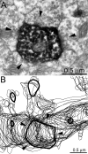
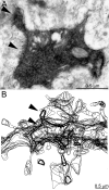
GABA-negative synapses (arrowheads) with spines of granule cell basal dendrites in epileptic rats. A Electron micrograph of basal dendrite #1 and its detached spines (in this section) labeled with electron dense reaction product. B Reconstructed segment of basal dendrite #1. The bold contours are of the basal dendrite profile shown in A. This region corresponds to cyan box #5AB in Figure 3C1. C Electron micrograph of basal dendrite #2 labeled with electron dense reaction product, which is lighter in the spine head than in the shaft. Within the field of view a GABA-positive axon, labeled by 10-nm-diameter black colloidal-gold particles, synapses with a GABA-negative shaft (arrow). D Reconstructed segment of basal dendrite. The bold contour is of the basal dendrite profile shown in C. This region corresponds to box #5CD in Figure 3F1.
Figure 6
Figure 6
GABA-negative and GABA-positive axon terminals synapse (arrowheads) with the same spine of granule cell basal dendrites in epileptic rats. A Electron micrograph of a spine of basal dendrite #1 labeled with electron dense reaction product. GABA-immunoreactivity is indicated by small black particles, which are 10-nm-iameter colloidal gold. B Reconstructed segment of basal dendrite #1. The bold contours are of the basal dendrite profile shown in A. This region corresponds to cyan box #6AB in Figure 3C1. C Electron micrographs of basal dendrite #2 labeled with electron dense reaction product, which is lighter in the spine head than in the shaft. GABA-immunoreactivity is indicated by small black particles, which are 10-nm-diameter colloidal gold. D Reconstructed segment of basal dendrite. The bold contours are of the basal dendrite profile shown in C. This region corresponds to box #6CD in Figure 3F1.
Figure 7
Figure 7
Synapses (arrowheads) with a spine and shaft, including a GABA-positive synapse with the shaft of a granule cell basal dendrite in an epileptic rat. A Electron micrograph. The basal dendrite and its detached spine (in this section) are labeled with electron dense reaction product. GABA-immunoreactivity is indicated by small black particles, which are 10-nm-diameter colloidal gold. Within the field of view a GABA-negative axon synapses with a GABA-positive dendritic shaft (arrow). B Reconstructed segment of basal dendrite. The bold contours are of the basal dendrite profile shown in A. This region corresponds to cyan box #7 in Figure 3C1.
Figure 8
Figure 8
Multiple GABA-negative synapses (arrowheads) with the shaft of a granule cell basal dendrite in an epileptic rat. A Electron micrograph. The basal dendrite is labeled with electron dense reaction product. B Reconstructed segment of basal dendrite. The bold contours are of the basal dendrite profile shown in A. This region corresponds to cyan box #8 in Figure 3C1.
Figure 9
Figure 9
GABA-negative synapses (arrowheads) with a spine and shaft of a granule cell basal dendrite in an epileptic rat. A Electron micrograph. The basal dendrite and its spine are labeled with electron dense reaction product, which is lighter in the spine head. B Reconstructed segment of basal dendrite. The bold contours are of the basal dendrite profile shown in A. This region corresponds to box #9 in Figure 3F1.
Figure 10
Figure 10
Synapses (arrowheads) between a GABA-negative axon and a spine and between a GABA-positive axon and shaft of a granule cell basal dendrite in an epileptic rat. A Electron micrograph. The basal dendrite and its spine are labeled with electron dense reaction product. GABA-immunoreactivity is indicated by small black particles, which are 10-nm-diameter colloidal gold. B Reconstructed segment of basal dendrite. The bold contours are of the basal dendrite profile shown in A. This region corresponds to box #10 in Figure 3F1.
Figure 11
Figure 11
Summary of synapses with granule cell basal dendrites #1 and #2 from epileptic rats. Proximal is up, distal is down. Synapses are indicated by markers. Most synapses are with GABA-negative spines. “Immuno.-unk.” synapses of basal dendrite #1 were in tissue sections that were used for the reconstruction but were not processed for immunocytochemistry.

Similar articles

Cited by

References

    1. Al-Hussain S, Al-Ali S. A Golgi study of cell types in the dentate gyrus of the adult human brain. Cell Mole Neurobiol. 1995;15:207–220. - PMC - PubMed
    1. André V, Marescaux C, Nehlig A, Fritschy JM. Alterations of hippocampal GABAergic system contribute to development of spontaneous recurrent seizures in the rat lithium-pilocarpine model of temporal lobe epilepsy. Hippocampus. 2001;11:452–468. - PubMed
    1. Austin JE, Buckmaster PS. Recurrent excitation of granule cells with basal dendrites and low interneuron density and inhibitory postsynaptic current frequency in the dentate gyrus of macaque monkeys. J Comp Neurol. 2004;476:205–218. - PubMed
    1. Babb TL, Pretorius JK, Kupfer WR, Crandall PH. Glutamate decarboxylase-immunoreactive neurons are preserved in human epileptic hippocampus. J Neurosci. 1989;9:2562–2574. - PMC - PubMed
    1. Bausch SB, Chavkin C. Changes in hippocampal circuitry after pilocarpine-induced seizures as revealed by opioid receptor distribution and activation. J Neurosci. 1997;17:477–492. - PMC - PubMed

Publication types

MeSH terms