

doi: 10.1021/ja802214x.
Epub 2008 May 8.
Structure of a 129Xe-cryptophane biosensor complexed with human carbonic anhydrase II
Affiliations
- PMID: 18461940
- PMCID: PMC2408383
- DOI: 10.1021/ja802214x
Item in Clipboard
Structure of a 129Xe-cryptophane biosensor complexed with human carbonic anhydrase II
J Am Chem Soc.
.
Abstract
Cryptophanes represent an exciting class of xenon-encapsulating molecules that can be exploited as probes for nuclear magnetic resonance imaging. The 1.70 A resolution crystal structure of a cryptophane-derivatized benezenesulfonamide complexed with human carbonic anhydrase II shows how an encapsulated xenon atom can be directed to a specific biological target. The crystal structure confirms binding measurements indicating that the cryptophane cage does not strongly interact with the surface of the protein, which may enhance the sensitivity of 129Xe NMR spectroscopic measurements in solution.
Figures
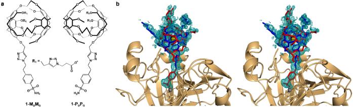
(a) The MoMo and PoPo enantiomers of the cryptophane-A-derived CA biosensor. The benzenesulfonamide moiety serves as an affinity tag that targets the Zn2+ ion, and the R1 substituents contain triazole propionate moieties that enhance aqueous solubility. (b) Stereoview of a simulated annealing omit map showing 1-MoMo (blue) and 1-PoPo (red) bound in the active site (1.9 σ contour, teal). A Bijvoet difference Fourier map (2.0 σ, black) confirms the encapsulation of Xe (yellow). Coordination interactions with Zn2+ (grey sphere) are indicated by dotted lines.
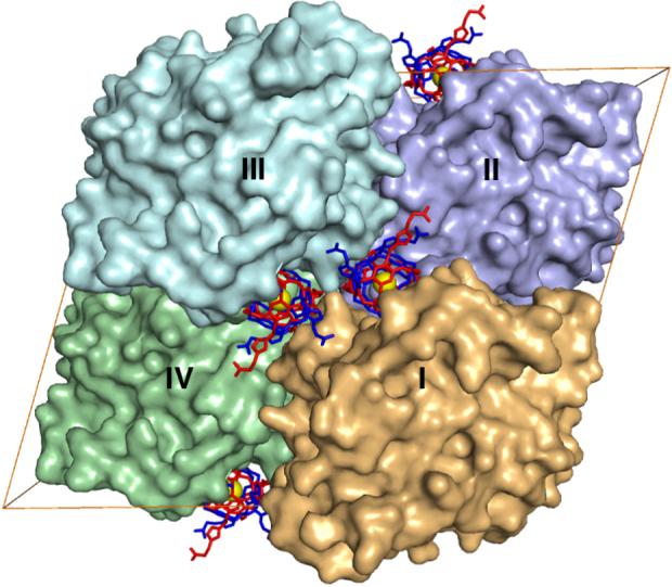
The unit cell of CAII crystals in space group C2 contains four molecules: I (x,y,z), II (x+1/2,y+1/2,z), III (-x,y,-z) and IV (-x+1/2,y+1/2,-z). The binding of 1 in the active site cleft of molecule I buries ∼500 Å2. Crystal contacts bury an additional 540 Å2 of the surface of 1 as follows: 270 Å2 with molecule III, and 240 Å2 and 30 Å2 with the front and back faces of molecule II, respectively. Molecule IV does not contact 1 bound to molecule I.
References
-
- Supuran CT, Scozzafava A. Bioorg. Med. Chem. 2007;15:4336–4350. - PubMed
-
- Elbaum D, Nair SK, Patchan MW, Thompson RB, Christianson DWJ. Am. Chem. Soc. 1996;118:8381–8387.
-
- Bozym RA, Thompson RB, Stoddard AK, Fierke CA. ACS Chem. Biol. 2006;1:103–111. - PubMed
-
- Cherubini A, Bifone A. Prog. Nucl. Magn. Reson. Spectrosc. 2003;42:1–30.
Publication types
MeSH terms
Substances
Grants and funding
LinkOut - more resources
Full Text Sources
Molecular Biology Databases
