. 2008 May 6;6(5):e105.
doi: 10.1371/journal.pbio.0060105.

Biogenesis of the trypanosome endo-exocytotic organelle is cytoskeleton mediated

Affiliations

Biogenesis of the trypanosome endo-exocytotic organelle is cytoskeleton mediated

Mélanie Bonhivers et al. PLoS Biol. .

Abstract

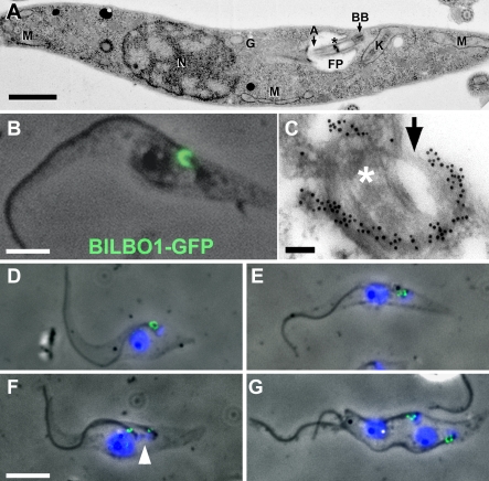
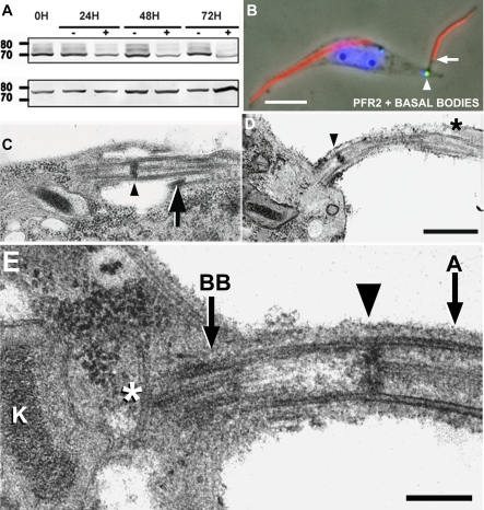
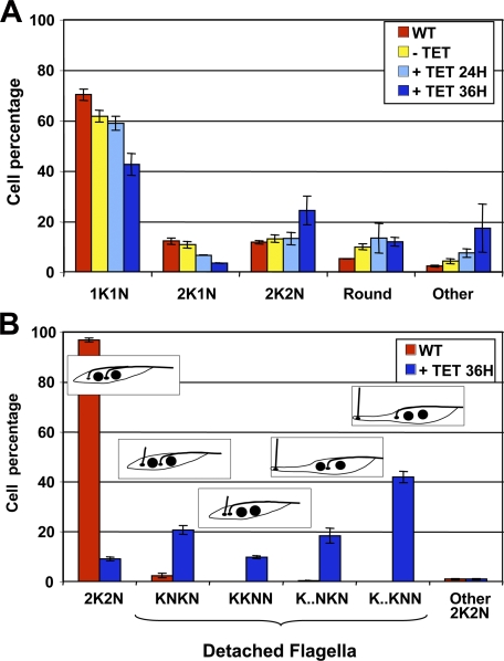
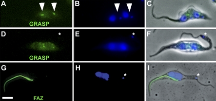
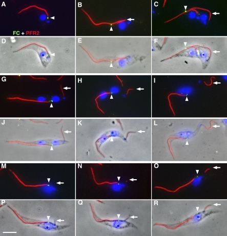
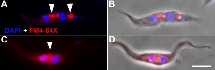
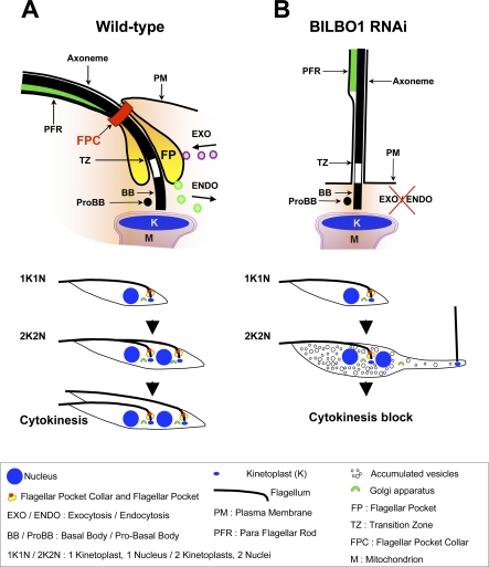
Trypanosoma brucei is a protozoan parasite that is used as a model organism to study such biological phenomena as gene expression, protein trafficking, and cytoskeletal biogenesis. In T. brucei, endocytosis and exocytosis occur exclusively through a sequestered organelle called the flagellar pocket (FP), an invagination of the pellicular membrane. The pocket is the sole site for specific receptors thus maintaining them inaccessible to components of the innate immune system of the mammalian host. The FP is also responsible for the sorting of protective parasite glycoproteins targeted to, or recycling from, the pellicular membrane, and for the removal of host antibodies from the cell surface. Here, we describe the first characterisation of a flagellar pocket cytoskeletal protein, BILBO1. BILBO1 functions to form a cytoskeleton framework upon which the FP is made and which is also required and essential for FP biogenesis and cell survival. Remarkably, RNA interference (RNAi)-mediated ablation of BILBO1 in insect procyclic-form parasites prevents FP biogenesis and induces vesicle accumulation, Golgi swelling, the aberrant repositioning of the new flagellum, and cell death. Cultured bloodstream-form parasites are also nonviable when subjected to BILBO1 RNAi. These results provide the first molecular evidence for cytoskeletally mediated FP biogenesis.


Conflict of interest statement

Competing interests. The authors have declared that no competing interests exist.

Figures

Figure 1
Figure 1. BILBO1 Localisation and FPC Biogenesis
(A) A thin-section electron micrograph of a T. brucei PF cell. The image shows the structural characteristics of a 1K1N cell. Note; only trans-Golgi vesicles are observed, as the Golgi itself is not in the plane of the section. Asterisk (*) denotes the flagellum transition zone. Scale bar indicates 1 μm. A. axoneme; BB, basal body; G, Golgi; K, kinetoplast; M, mitochondria; N, nucleus. (B) Phase/fluorescence-micrograph of a procyclic cytoskeleton expressing BILBO1-eGFP. BILBO1-eGFP protein localises to the FPC. Scale bar indicates 2.5 μm. (C) Electron micrograph of a thin-sectioned WT cytoskeleton probed with anti-BILBO1 antiserum, followed by immunogold labelling. This illustrates the precise location of BILBO1 on the FPC. Asterisk (*) denotes the flagellum transition zone. Arrow denotes an unlabelled portion of the FPC. Scale bar indicates 100 nm. (D–G) Immunofluorescence of cytoskeletons probed with anti-BILBO1 antibody and counterstained with DAPI showing the FPC label and the duplication-segregation of the FPC during the cell cycle. Arrowhead in (F) denotes the kinetoplast, which has not completed S phase, and illustrates that the FPC is duplicated prior to kinetoplast S phase completion. Scale bar in (D–G) indicates 5 μm.
Figure 2
Figure 2. BILBO1 RNAi Prevents FP Biogenesis
(A). Western blot of BILBO1 RNAi-induced cells (+) probed with anti-BILBO1 antibody (upper panel) or control antibody L8C4 (anti-PFR2), 0–72 h of BILBO1 RNAi induction. This blot shows that BILBO1 protein levels diminish over time but are still present after 72 h of RNAi induction. (B) A phase-DAPI-immunofluorescence micrograph of a BILBO1 RNAi-induced 2K2N cytoskeleton probed with anti-PFR2 (L8C4) and anti-basal bodies (BBA4) (36-h induction). Basal body and kinetoplast are indicated by the arrowhead; the flagellum and site of PFR initiation are indicated by the arrow. Scale bar indicates 5 μm. (C) A micrograph of a thin section of the FP of a noninduced PF cell illustrating the transition zone (arrowhead) and the FPC (arrow). (D) A micrograph of BILBO1 RNAi-induced cell (48 h) at the new flagellum region. Note loss of flagellum-to-cell body attachment, PFR (asterisk), transition zone location (arrowhead), and absence of a FP. Scale bar indicates 500 nm. (E) Thin-section micrograph of BILBO1 RNAi-induced (48 h) cell illustrating the proximal end of the new flagellum and the absence of a FP. A portion of the basal body (BB) is located in the cell, whereas the transition zone (arrowhead) is external to the cell body. Note the presence of cytoplasmic microtubule(s) (absent in WT cells) at the proximal region of the basal body (asterisk). K, kinetoplast. Scale bar indicates 200 nm.
Figure 3
Figure 3. RNAi of BILBO1 Disrupts Cytokinesis
(A) WT and BILBO1 RNAi PF cells were scored for kinetoplast/nuclei by DAPI labelling at 0, 24 h, and 36 h postinduction (+ TET). After 36 h of induction, a large number of 2K2N cell types are produced, and the proportion of the 2K1N cell type diminishes significantly. Induced cells arrest in the 2K2N configuration. The “Round” category represents round PF cells in which neither the nucleus nor the kinetoplasts could be individually distinguished. The “Other” categories represent cells in which 2N or 2K could not be assessed. (B) The distribution of 2K2N cell types in WT and BILBO1 RNAi-induced cells (36 h). Cell morphology was scored by phase contrast microscopy and DAPI labelling as well as for the number of cells with flagella that had lost their flagellum-to-cell body attachment. The “Other” 2K2N category represents PF cells in which the position of the flagellum could not be assessed. Five distinctive 2K2N phenotypes were observed in induced PF cells: (1) 2K2N cells that appeared normal in kinetoplast and nuclear positioning (KNKN [8.96% SE ± 0.82%]); (2) KNKN cells with a loss of new flagellum-to-cell body attachment (20.56% SE ± 1.76%); (3) KKNN cells with a loss of new flagellum-to-cell body attachment (9.63% SE ± 0.63%); (4) elongated KNKN cells (18.33% SE ± 3.01%); and (5) elongated KKNN cells (41.73% SE ± 2.3%).
Figure 4
Figure 4. RNAi Knockdown of BILBO1 Induces Loss of Basal Body–Mediated Golgi Segregation and Causes Defects of Important Cytoskeletal Structures
(A–C) A nontransformed 2K2N cell probed with anti-GRASP (green) and DAPI (blue), illustrating two major GRASP signals (arrowheads) located between the segregated kinetoplasts and nuclei. (D–F) A BILBO1 RNAi-induced (36 h) 2K2N cell probed with anti-GRASP. The two GRASP signals are observed near the nuclei. Despite a limited degree of Golgi segregation, no GRASP signal is observed near the new kinetoplast. The kinetoplast and the new flagellum (asterisk) are located in the extreme posterior end of the cell. (G–I) BILBO1 and the FPC are important for cytoskeleton organisation. Immunofluorescence micrograph of a PF cytoskeleton probed with L3B2 (anti-FAZ) antibody after BILBO1 RNAi knockdown (36 h). The flagellum-to-cell body attachment is lost, and the new flagellum is located at the posterior region of the cell. No new FAZ is formed, whereas the old FAZ remains associated with the old flagellum. The kinetoplast (asterisk) is located in the extreme posterior of the cell. Scale bar indicates 5 μm.
Figure 5
Figure 5. The New Flagellum of BILBO1 RNAi-Induced Cells Loses Physical Contact with the Old Flagellum Early in the Cell Cycle
(A–F) Uninduced BILBO1 RNAi cytoskeletons probed with DAPI and double-labelled with anti-Flagellum Connector AB1 (green) and anti-PFR2 L8C4 (red) antibodies showing the attachment of the new flagellum to the maternal old flagellum and movement of the FC with the growth of the new flagellum. (A and D) Merged images of a 1K1N cytoskeleton in which the FC is located in the FP of the old flagellum. (B and E) Merged images of a 2K1N2F (two kinetoplasts, one nucleus, and two flagella) cytoskeleton in which the FC is present at the distal end of the new flagellum. (C and F) Merged images of a 2K2N2F postmitotic cytoskeleton in which the FC is present at the distal end of the new flagellum. (D, E, and F) are phase contrast merged images of (A, B, and C), respectively. (G–L) 2K2N2F-induced BILBO1 RNAi cytoskeletons showing the extended posterior end of the cell and the new flagellum-to-cell body attachment is disrupted. The FC is present only in the FP of the old flagellum. (J, K, and L) are phase contrast merged images of (G, H, and I), respectively. (M–R) 2K1N2F BILBO1 RNAi-induced cytoskeletons showing early stages in the phenotype in which the new flagellum is clearly not attached to the old flagellum. The FC is present only in the FP of the old flagellum. In (M and P), no PFR2 signal is detectable on the new flagellum. This new flagellum is at a very early stage of growth; it is not attached to the old flagellum and has a detached flagellum-to-cell body phenotype. The FC remains in the FP. (P, Q, and R) are phase contrast merges of (M, N, and O) respectively. Arrowheads denote the FC signal, and arrows denote the new flagellum. Scale bar indicates 5 μm.
Figure 6
Figure 6. No Endocytotic Activity Is Associated with the New Flagellum
A 2K2N WT cell (A and B) and a BILBO1 RNAi-induced cell for 36 h (C and D) were labelled with DAPI (blue) and the red fluorescent lipophilic dye FM4-64X (red). Arrowheads in (A) denote areas of endocytotic activity associated with both FPs of this cell. In (C), a large area of endocytotic activity, in the region of the old FP, is labelled, but no activity is associated with the new flagellum at the posterior end of the cell. (B and D) are DAPI-phase-fluorescence merged images of (A and C). Scale bar indicates 5 μm.
Figure 7
Figure 7. A Schematic Diagram That Describes the Normal Procyclic T. brucei Cell Division Cycle and the Fate of Induced BILBO1 RNAi Cells
This schematic shows the organisation of the flagellum, function of the FP, and location of the FPC in a WT cell (A). The lower half of (A) illustrates the normal division cycle of the FP, Golgi, and flagellum in WT cells, whereas the drastically altered morphology of an induced BILBO1 RNAi cell (B) shows the absence of a FP and the loss of new flagellum-to-cell body attachment. The lower half of (B) shows an example of the major phenotype formed resulting from BILBO1 RNAi knockdown in procyclic cells. BILBO1 RNAi prevents FPC and FP biogenesis, disrupts endo-exocytosis, initiates cytokinesis block, and induces new flagellum cell body detachment and relocation to the posterior of the cell.

Similar articles

Cited by

References

    1. Landfear SM, Ignatushchenko M. The flagellum and flagellar pocket of trypanosomatids. Mol Biochem Parasitol. 2001;115:1–17. - PubMed
    1. Overath P, Engstler M. Endocytosis, membrane recycling and sorting of GPI-anchored proteins: Trypanosoma brucei as a model system. Mol Microbiol. 2004;53:735–744. - PubMed
    1. De Souza W. Secretory organelles of pathogenic protozoa. An Acad Bras Cienc. 2006;78:271–291. - PubMed
    1. Garcia-Salcedo JA, Perez-Morga D, Gijon P, Dilbeck V, Pays E, et al. A differential role for actin during the life cycle of Trypanosoma brucei. EMBO J. 2004;23:780–789. - PMC - PubMed
    1. Field MC, Natesan SK, Gabernet-Castello C, Lila Koumandou V. Intracellular trafficking in the trypanosomatids. Traffic. 2007;8:629–639. - PubMed

Publication types

Substances

Associated data