. 2008 Jun 15;318(2):258-67.
doi: 10.1016/j.ydbio.2008.03.022. Epub 2008 Mar 26.

Loss of unc45a precipitates arteriovenous shunting in the aortic arches

Affiliations

Loss of unc45a precipitates arteriovenous shunting in the aortic arches

Matthew J Anderson et al. Dev Biol. .

Abstract

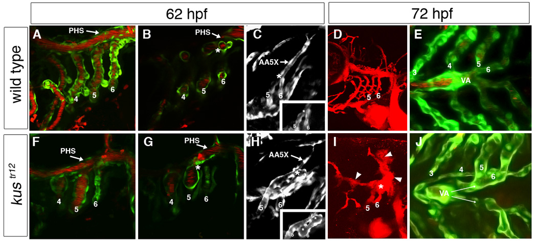
Aortic arch malformations are common congenital disorders that are frequently of unknown etiology. To gain insight into the factors that guide branchial aortic arch development, we examined the process by which these vessels assemble in wild type zebrafish embryos and in kurzschluss(tr12) (kus(tr12)) mutants. In wild type embryos, each branchial aortic arch first appears as an island of angioblasts in the lateral pharyngeal mesoderm, then elaborates by angiogenesis to connect to the lateral dorsal aorta and ventral aorta. In kus(tr12) mutants, angioblast formation and initial sprouting are normal, but aortic arches 5 and 6 fail to form a lumenized connection to the lateral dorsal aorta. Blood enters these blind-ending vessels from the ventral aorta, distending the arteries and precipitating fusion with an adjacent vein. This arteriovenous malformation (AVM), which shunts nearly all blood directly back to the heart, is not exclusively genetically programmed, as its formation correlates with blood flow and aortic arch enlargement. By positional cloning, we have identified a nonsense mutation in unc45a in kus(tr12) mutants. Our results are the first to ascribe a role for Unc45a, a putative myosin chaperone, in vertebrate development, and identify a novel mechanism by which an AVM can form.


Figures

Fig. 1
Fig. 1. Branchial aortic arch and ventral aorta development
Aortic arches 3–6 first appear in an anterior-to-posterior sequence as angioblast islands (indicated as numbers 3 through 6) in the lateral pharyngeal mesoderm; sprouts emerge from these islands and travel ventromedially and dorsally (A–L; see also Movie 1). By 48 hpf (M), ventral sprouts merge to form bilateral ventral aortae (VA), which fuse into a single midline vessel by 72 hpf (N). Dorsally (O; 72 hpf), branchial aortic arches turn medially beneath the primary head sinus (PHS). Aortic arches 3 and 4 connect independently to the lateral dorsal aortae, whereas aortic arches 5 and 6 merge and extend to the lateral dorsal aortae as a single vessel, the aortic arch 5 extension (AA5X). A–D: tie1 in situ hybridization. E–H and N: 2D confocal projections of TG(flk1:GFPla116;gata1:dsRed) embryos, labeling endothelial and blood cells green and red, respectively. E–H are representative time points taken from Movie 1. M,O: 2D confocal projections of TG(flk1:GFPla116) embryos. I–L: wiring diagrams of images in E–H; vessels were traced using Photoshop. A–L: lateral views, anterior to the left. M,N: ventral views, anterior to the left. O: dorsal view, anterior to the left.
Fig. 2
Fig. 2. Aortic arches 5 and 6 exhibit delayed lumenization and fail to properly connect to the lateral dorsal aortae in kustr12 mutants
In wild type embryos, aortic arches 5 and 6 are lumenized by 50 hpf (A) and exhibit strong blood flow by 52 hpf (C). In kustr12 (B,D), lumenization is delayed by 2–3 hours and blood cells accumulate in distal portions of aortic arch 5 (arrow). This accumulation of blood cells is accompanied by failure of the AA5X to make a patent connection to the LDA [compare E (WT) to F (kustr12)]. A–F, 2D confocal projections of TG(flk1:GFPla116; gata1:dsRed) embryos at 50 hpf (A,B) or 52 hpf (C–F), labeling endothelial and blood cells green and red, respectively. A–D, lateral views, anterior to the left. E–F, dorsal views, anterior to the left.
Fig. 3
Fig. 3. Arteriovenous malformation and ventral aorta defect in kustr12 mutants
In wild type embryos, aortic arches 5 and 6 fuse just behind the primary head sinus (PHS) and connect to the lateral dorsal aorta via an extension of aortic arch 5 (AA5X); clear demarcation can be seen between the aortic arch 5/6 connection (asterisk) and the PHS (A–C). In contrast, in kustr12 mutants, AA5X is poorly formed, and the enlarged aortic arch 5/6 connection fuses to the adjacent PHS (asterisk), creating an AVM (arrowhead) that shunts blood directly back to the heart (F–H). In addition, the AA5X is poorly formed (H). This AVM (asterisk) carries the majority of blood flow and is typically associated with localized hemorrhage [arrowheads; compare D (WT) to I (kustr12)]. In wild type embryos, the paired ventral aortae (VA) fuse by 72 hpf (E). In contrast, in kustr12, the ventral aortae fail to fuse properly and remain paired at 72 hpf, similar to wild types at 48 hpf (J; compare also to Fig. 1M). 2D confocal projections of TG(flk1:GFPla116;gata1:dsRed) embryos (A,E,F,J); TG(flk1:GFPla116) embryos (C,H); or microangiograms (D,I). B, G and insets in C,H are single planes from confocal Z-stacks represented by A,F,C, and H, respectively. A–C; F–H: 62 hpf. D,E,I,J: 72 hpf. A, B, D, F, G, I: lateral views, anterior to the left. C,H: dorsolateral views, anterior to the left. E,J: ventral views, anterior to the left.
Fig. 4
Fig. 4. Increased distance between left and right posterior ceratobranchial cartilages is a specific kustr12 phenotype
Aortic arches (bright green) develop in close association with pharyngeal cartilages (red) in WT (A) and kustr12 (D). Note the malformed ventral aorta (VA) in kustr12. (The darker green staining is background and represents pharyngeal muscle.) Defects in jaw and pharyngeal cartilages in kustr12 include shortening of the palatoquadrates (PQ); compression of the basibranchial cartilages (horizontal white line); improper angling of the ceratobranchial (asterisks) and ceratohyal (arrowheads) cartilages; and decreased lateral distance (vertical white lines) between left and right ceratobranchial cartilages in pharyngeal arches 5 and 6 [compare B (WT) to E (kustr12)]. Like kustr12, vbgy6 mutants (C) and tnnt2 morphants (F) develop edema and exhibit all described cartilage defects except increased lateral distance between posterior ceratobranchials. Thus, only the latter defect is likely a direct effect of the kustr12 mutation. A,D: 2D confocal projections of TG(flk1:GFPla116; gata1:dsRed) embryos stained with anti-collagen II (red) and anti-GFP (green). B,C,E,F: Macro images of embryos stained with anti-collagen II. All embryos are 4 dpf; ventral views, anterior to the left.
Fig. 5
Fig. 5. Lack of blood flow can rescue AVM formation in kustr12 mutants
Ninety-eight percent (n = 117) of wild type or heterozygous embryos treated with 2,3-butanedione monoxime (BDM; stops heart beat) between 47 and 53 hpf exhibited normal aortic arches (A), ventral aorta (C), and jaw and pharyngeal cartilages (E). This treatment rescued aortic arch (B) and ventral aorta (D) phenotypes in 67% of kustr12 mutants (n = 29), but had no effect on the basibranchial cartilage phenotype (vertical lines in F). A–D: 2D confocal projections of 74 hpf TG(flk1:GFPla116;gata1:dsRed) embryos, labeling endothelial and blood cells green and red, respectively. G–H: Macro images of 4 dpf embryos stained with anti-collagen II. A,B: lateral views, anterior the left. C,D,E,F: ventral views, anterior to the left. PHS, primary head sinus. VA, ventral aorta.
Fig. 6
Fig. 6. unc45a is mutated in kustr12 mutants
(A) Fine mapping placed kustr12 in a 0.09 cM interval on chromosome 25 (blue bar), which contains unc45a (gray shading). (B) Unc45a contains an N-terminal TPR domain, a central domain of unknown function, a C-terminal UCS domain, and armadillo (ARM) repeats. The protein is encoded by 20 exons. (C) Phylogenetic analysis clearly indicates that this gene is homologous to mammalian unc45a, and not the closely related gene, unc45b, which is muscle-specific. (D) Sequencing of unc45a cDNAs in kustr12 mutants and wild type siblings revealed a T-to-A transversion in unc45a, converting a leucine to a stop codon. This mutation truncates the protein within exon 14 (B), deleting the armadillo repeats and a large portion of the UCS domain.
Fig. 7
Fig. 7. Rescue and phenocopy experiments confirm the role of unc45a in the kustr12 vascular phenotype
Injection of full length (B; n = 90) but not C-terminally truncated (C; n = 13) unc45a mRNA (unc45akus, corresponding to the kustr12 allele) restored a wild type vascular phenotype to 67% of kustr12 mutants (compare to uninjected kustr12, A). Conversely, injection of am unc45a translation blocking morpholino (trans MO, E; n = 144) or exon 13/14 splice donor site blocking morpholino (splice MO, F; n = 60) produced a kustr12-like AVM phenotype in 20% and 23%, respectively, of wild type embryos (compare to uninjected wild type, D). A–C, F: 2D confocal projections of 74 hpf TG(flk1:GFPla116;gata1:dsRed) embryos, labeling endothelial cells green and blood cells red. D–E: 2D confocal projections of angiograms of 74 hpf embryos. Lateral views, anterior to the left. PHS, primary head sinus.
Fig. 8
Fig. 8. Developmental expression patterns of unc45a mRNA
In situ hybridization reveals diffuse, ubiquitous unc45a expression at 64-cell (A) and shield stage (B). By the three somite stage (C), there is specific expression in the polster (p). At 24 hpf (D), additional expression domains arise, including retina (r), midbrain (m), and hindbrain (h). By 46 hpf (E–G), expression continues in retina and brain, and is also strong in the liver (l, and arrow in E inset), pharynx (ph), gut (not shown), and pharyngeal arches (pa). The mosaic expression pattern in the pharyngeal arches is illustrated in G. This general pattern continues up to 5 dpf (H), with expression becoming more laterally restricted and segmented in the pharyngeal arches. A–B: animal pole at top. C–E, H: lateral views, anterior to the left. Inset in E: dorsal view, anterior to the left. F: transverse section. G: coronal section.

References

    1. Ao W, Pilgrim D. Caenorhabditis elegans UNC-45 is a component of muscle thick filaments and colocalizes with myosin heavy chain B, but not myosin heavy chain A. J Cell Biol. 2000;148:375–384. - PMC - PubMed
    1. Barral JM, et al. Unc-45 mutations in Caenorhabditis elegans implicate a CRO1/She4p-like domain in myosin assembly. J Cell Biol. 1998;143:1215–1225. - PMC - PubMed
    1. Barral JM, et al. Role of the myosin assembly protein UNC-45 as a molecular chaperone for myosin. Science. 2002;295:669–671. - PubMed
    1. Bazzaro M, et al. Myosin II Co-Chaperone General Cell UNC-45 Overexpression Is Associated with Ovarian Cancer, Rapid Proliferation, and Motility. Am J Pathol. 2007;171:1640–1649. - PMC - PubMed
    1. Blatch GL, Lassle M. The tetratricopeptide repeat: a structural motif mediating protein-protein interactions. Bioessays. 1999;21:932–939. - PubMed

Publication types

MeSH terms