. 2008 Jun 27;283(26):17753-6.
doi: 10.1074/jbc.C800040200. Epub 2008 May 6.

Crystal structure of squid rhodopsin with intracellularly extended cytoplasmic region

Affiliations

Crystal structure of squid rhodopsin with intracellularly extended cytoplasmic region

Tatsuro Shimamura et al. J Biol Chem. .

Abstract

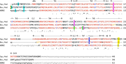
G-protein-coupled receptors play a key step in cellular signal transduction cascades by transducing various extracellular signals via G-proteins. Rhodopsin is a prototypical G-protein-coupled receptor involved in the retinal visual signaling cascade. We determined the structure of squid rhodopsin at 3.7A resolution, which transduces signals through the G(q) protein to the phosphoinositol cascade. The structure showed seven transmembrane helices and an amphipathic helix H8 has similar geometry to structures from bovine rhodopsin, coupling to G(t), and human beta(2)-adrenergic receptor, coupling to G(s). Notably, squid rhodopsin contains a well structured cytoplasmic region involved in the interaction with G-proteins, and this region is flexible or disordered in bovine rhodopsin and human beta(2)-adrenergic receptor. The transmembrane helices 5 and 6 are longer and extrude into the cytoplasm. The distal C-terminal tail contains a short hydrophilic alpha-helix CH after the palmitoylated cysteine residues. The residues in the distal C-terminal tail interact with the neighboring residues in the second cytoplasmic loop, the extruded transmembrane helices 5 and 6, and the short helix H8. Additionally, the Tyr-111, Asn-87, and Asn-185 residues are located within hydrogen-bonding distances from the nitrogen atom of the Schiff base.


Figures

FIGURE 1.
FIGURE 1.
Structural sequence alignment of squid rhodopsin, bovine rhodopsin and β2AR. The structural alignment was based on the 3D-Coffee alignment (4). Residues on helix regions are colored red, and residue(s) of helix bending are colored in blue. Transmembrane helical regions (TH1–TH7) and helix H8 with extracellular (EL1–EL3) and cytoplasmic (CL1–CL3) loops, and each helix in N- and C-terminal tails (NH and CH) are indicated. Posttranslational modifications are shaded by the following colors: cyan, N-glycosylation; a pair of pink or green, disulfide bridge(s); yellow, palmitoylated cysteine; blue, Schiff-based lysine with 11-cis-retinal; gold, N-terminal methionine acetylation. Residues indicated by small letters are not in models but in crystal protein samples, and residues indicated by small letters in italic gray do not exist in the crystal sample proteins due to expression processing, protease digestion, or protein engineering. Squ_rhod, squid rhodopsin (PDB code: 2ZIY in this study); Bov_rhod, bovine rhodopsin (1F88 (5) or 1GZM (17)); and ADRB2 (2RH1 (8)).
FIGURE 2.
FIGURE 2.
Crystal structure of squid rhodopsin. A, schematic model of squid rhodopsin with multicolored cylindrical helices. Transmembrane helices are indicated as TH1–TH7, and amphipathic short helix H8 is indicated as H8. 11-cis-Retinal at Lys-305 and palmitoylated cysteines Cys-336 and Cys-337 are indicated by the yellow sphere-and-stick models. A hydrophilic short helix in each N- and C-terminal tail is indicated as NH and CH, respectively. N and C termini are indicated by the letters N and C, and the cytoplasmic loop 3 is indicated as CL3. Putative transmembrane-spanning regions are indicated by yellow belts. B, superimposed schematic models and electrostatic surfaces of known GPCR structures. Superimposed schematic structures of squid and bovine rhodopsins, and β2AR are shown. Squid rhodopsin in this study is shown in orange, bovine rhodopsin in the trigonal crystal (1GZM) (17) is blue, and β2AR (2RH1) excluding the T4 lysozyme part of CL3 (8) is sky blue. The electrostatic surfaces of squid rhodopsin (Squ Rhod), bovine rhodopsin in the trigonal crystal (Bov Rhod), and β2AR (β2AR) are represented in blue (positive) to red (negative) with the squid rhodopsin structure in an orange schematic. To clarify the contribution of the distal C-terminal tail, the electrostatic surface of squid rhodopsin without the C-terminal tail after Glu-343 was also calculated (Squ C-trc). Different TH5 regions are indicated by the red line.

References

    1. Pierce, K. L., Premont, R. T., and Lefkowitz, R. J. (2002) Nat. Rev. Mol. Cell Biol. 3 639–650 - PubMed
    1. Elefsinioti, A. L., Bagos, P. G., Spyropoulos, I. C., and Hamodrakas, S. J. (2004) BMC Bioinformatics 5 208. - PMC - PubMed
    1. Sgourakis, N. G., Bagos, P. G., and Hamodrakas, S. J. (2005) Bioinformatics 21 4101–4106 - PubMed
    1. Poirot, O., Suhre, K., Abergel, C., O'Toole, E., and Notredame, C. (2004) Nucleic Acids Res. 32 W37–40 - PMC - PubMed
    1. Palczewski, K., Kumasaka, T., Hori., T., Behnke, C. A., Motoshima, H., Fox, B. A., Le Trong, I., Teller, D. C., Okada, T., Stenkamp, R. E., Yamamoto, M., and Miyano, M. (2000) Science 289 739–745 - PubMed

Associated data

LinkOut - more resources