. 2008 Aug;36(8):1637-49.
doi: 10.1124/dmd.107.019562. Epub 2008 May 7.

Pathways of carbamazepine bioactivation in vitro. III. The role of human cytochrome P450 enzymes in the formation of 2,3-dihydroxycarbamazepine

Affiliations

Pathways of carbamazepine bioactivation in vitro. III. The role of human cytochrome P450 enzymes in the formation of 2,3-dihydroxycarbamazepine

Robin E Pearce et al. Drug Metab Dispos. 2008 Aug.

Abstract

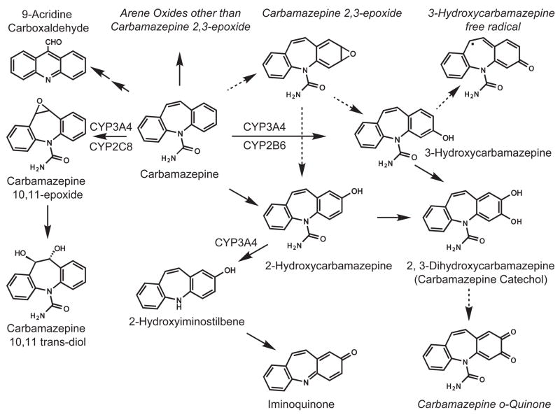
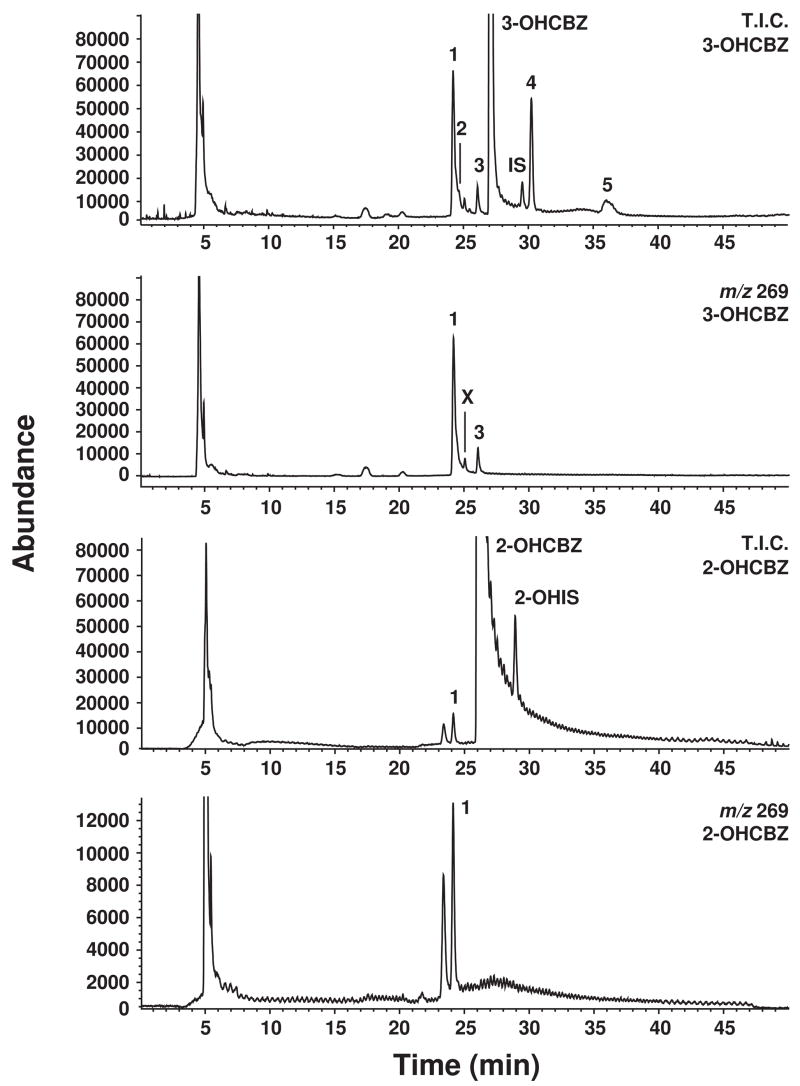
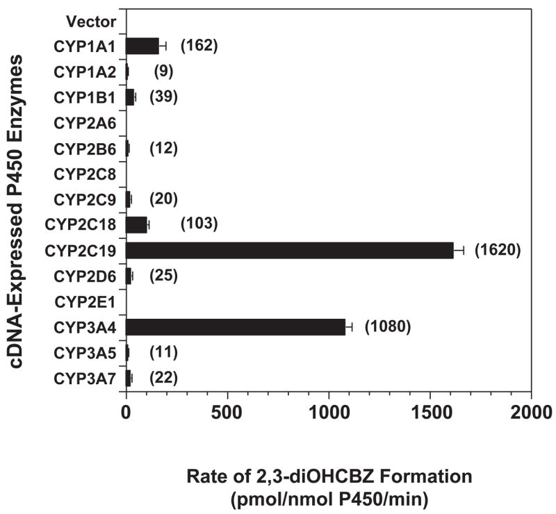
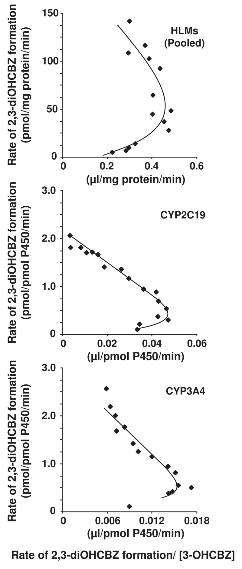
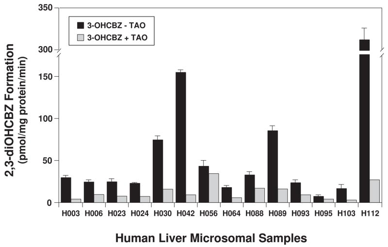
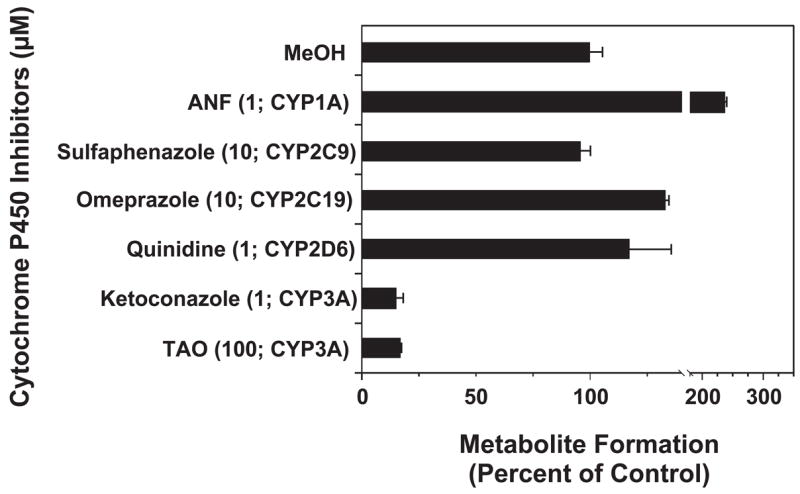
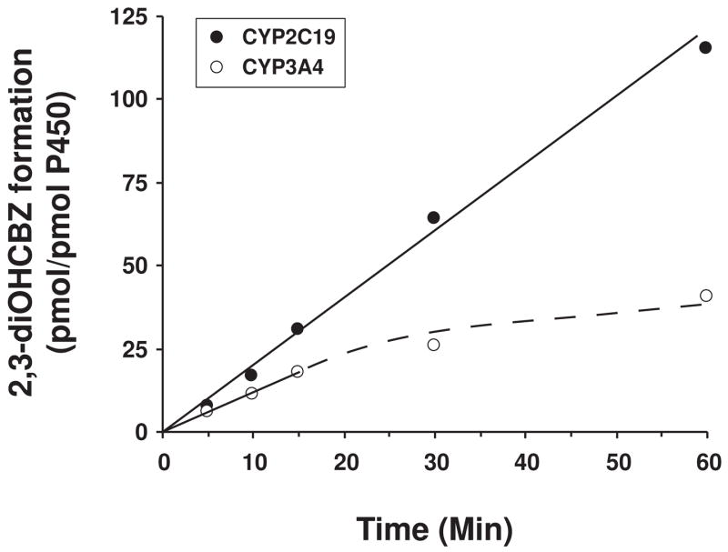
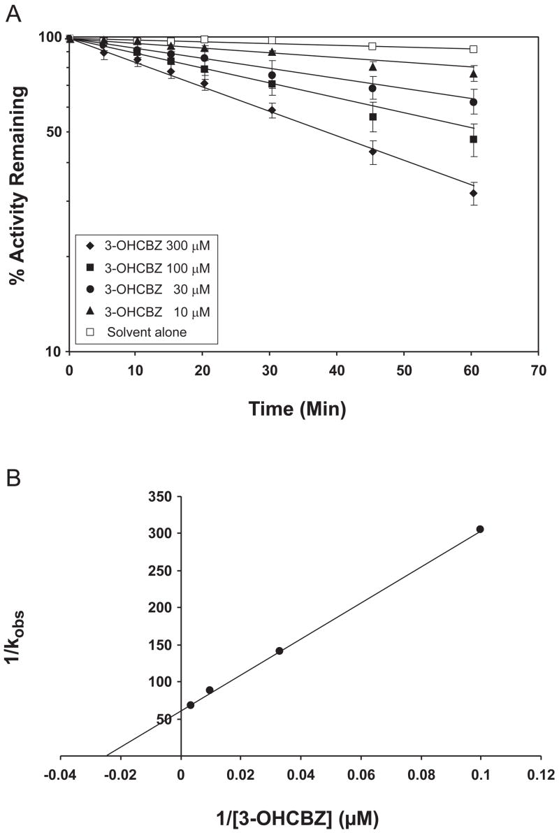
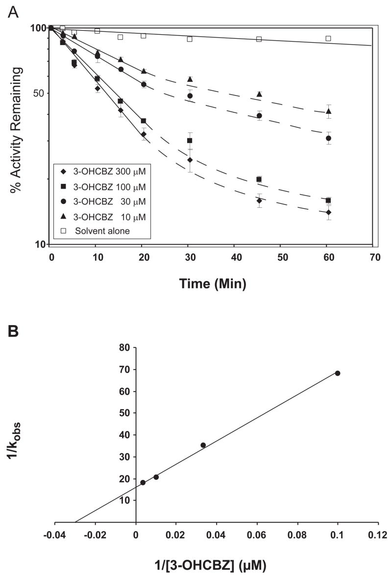
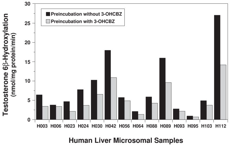
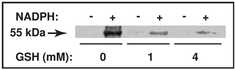
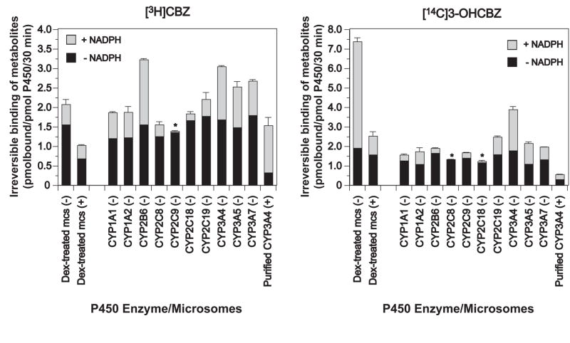
Conversion of the carbamazepine metabolite 3-hydroxycarbamazepine (3-OHCBZ) to the catechol 2,3-dihydroxycarbamazepine (2,3-diOHCBZ) followed by subsequent oxidation to a reactive o-quinone species has been proposed as a possible bioactivation pathway in the pathogenesis of carbamazepine-induced hypersensitivity. Initial in vitro phenotyping studies implicated CYP3A4 as a primary catalyst of 2,3-diOHCBZ formation: 2-hydroxylation of 3-OHCBZ correlated significantly (r(2) > or = 0.929, P < 0.001) with CYP3A4/5 activities in a panel of human liver microsomes (n = 14) and was markedly impaired by CYP3A inhibitors (>80%) but not by inhibitors of other cytochrome P450 enzymes (< or = 20%). However, in the presence of troleandomycin, the rate of 2,3-diOHCBZ formation correlated significantly with CYP2C19 activity (r(2) = 0.893, P < 0.001) in the panel of human liver microsomes. Studies with a panel of cDNA-expressed enzymes revealed that CYP2C19 and CYP3A4 were high (S50 = 30 microM) and low (S50 = 203 microM) affinity enzymes, respectively, for 2,3-diOHCBZ formation and suggested that CYP3A4, but not CYP2C19, might be inactivated by a metabolite formed from 3-OHCBZ. Subsequent experiments demonstrated that preincubation of 3-OHCBZ with human liver microsomes or recombinant CYP3A4 led to decreased CYP3A4 activity, which was both preincubation time- and concentration-dependent, but not inhibited by inclusion of glutathione or N-acetylcysteine. CYP3A4, CYP3A5, CYP3A7, CYP2C19, and CYP1A2 converted [14C]3-OHCBZ into protein-reactive metabolites, but CYP3A4 was the most catalytically active enzyme. The results of this study suggest that CYP3A4-dependent secondary oxidation of 3-OHCBZ represents a potential carbamazepine bioactivation pathway via formation of reactive metabolites capable of inactivating CYP3A4, potentially generating a neoantigen that may play a role in the etiology of carbamazepine-induced idiosyncratic toxicity.


Figures

Figure 1
Figure 1
Metabolic pathways and proposed reactive metabolites of CBZ. Italicized species are inferred from products.
Figure 2
Figure 2
Representative HPLC/MS chromatograms obtained by APCI at selected ion currents of [MH]+ ions of 3-OHCBZ or 2-OHCBZ and their respective metabolites formed by human liver microsomes. T.I.C., total ion count; 1, proposed 2,3-diOHCBZ metabolite (24.5 min); 2–5, unidentified metabolites of 3-OHCBZ (29.5 min); X, unidentified contaminant: IS, (CBZ, 29.5 min); 2-OHIS, 2hydroxyiminostilbene (28.5 min).
Figure 3
Figure 3
2,3-diOHCBZ formation by human recombinant cytochrome P450 enzymes. 3-OHCBZ (100 μM) was incubated with heterologously expressed human P450 enzymes as described under “Materials and Methods”. Each bar represents the mean ± S.D. of six determinations. No apparent formation of 2,3-diOHCBZ was catalyzed by microsomes containing the vector control or CYPs 2A6, 2C8 and 2E1. Formation of 2,3-diOHCBZ in microsomes containing the other cDNA-expressed human P450 enzymes examined was found to be statistically different from the limit of detection for 3-OHCBZ (a surrogate standard for 2,3-diOHCBZ; LOD = ~200 fmol) as determined by Student’s t test (p ≦ 0.05).
Figure 4
Figure 4
Effect of substrate concentration on the rate of 2,3-diOHCBZ formation by pooled human liver microsomes and recombinant CYP2C19 and CYP3A4 (Eadie-Hofstee plots). 3-OHCBZ (5–500 μM) was incubated with pooled human liver microsomes (0.05 mg microsomal protein) or microsomes from insect cells containing bacculovirus-expressed CYP2C19 or CYP3A4 (5 pmol) in 100-μl reaction mixtures at 37±0.1°C, and terminated with 100-μl of methanol. Incubations containing human liver micrsomes were terminated after 30 min, whereas incubations containing CYP3A4 and CYP2C19 were terminated after 10 and 15 min, respectively. Following precipitation of microsomal protein, an aliquot (75 μl) of the supernatant was analyzed by HPLC/MS via direct injection, respectively, as described under Materials and Methods.
Figure 5
Figure 5
Effect of troleandomycin (TAO) on the 2-hydroxylation of 3-OHCBZ by a panel of human liver microsomes. Human liver microsomes (0.05 mg microsomal protein) were incubated with 3-OHCBZ (100 μM) in 100-μl reaction mixtures at 37±0.1°C, terminated after 30 min with 100-μl of methanol and analyzed by HPLC/MS via direct injection, as described under Materials and Methods. Incubations containing the mechanism-based inhibitor TAO (100 μM) were pre-incubated with human liver microsomes and NADPH-generating system for 20 min before the reaction was started with substrate. Bars representing uninhibited rates of 2,3-diOHCBZ formation are the mean ± S.D. of six determinations, whereas bars representing rates of 2,3-diOHCBZ formation in the presence of TAO are the mean of duplicate determinations.
Figure 6
Figure 6
Effects of various P450 isoform-selective inhibitors on the formation of 2,3-diOHCBZ by human liver microsomes. Pooled human liver microsomes were incubated with 3-OHCBZ (100 μM) in the presence or absence of various chemicals, as described under “Materials and Methods”. Final inhibitor concentrations and the major P450 isoform inhibited are indicated in the brackets. Bars represent rates of 2,3-diOHCBZ formation (mean ± S.D. of duplicate determinations) and are expressed as a per cent of the control rate (96.8 ± 4.8 pmol/mg protein/min).
Figure 7
Figure 7
Effect of time on the formation of 2,3-diOHCBZ by human recombinant CYP2C19 and CYP3A4. 3-OHCBZ (100 μM) was incubated with insect cell microsomes containing baculovirus-expressed CYP2C19 or CYP3A4 (5 pmol/100-μl incubation) at 37±0.1°C for 0–60 min and analyzed by HPLC/MS as described under “Materials and Methods”. Each data point represents the mean of duplicate determinations.
Figure 8
Figure 8
Time- and concentration-dependent inactivation of testosterone 6β-hydroxylase activity in human liver microsomes. A. Human liver microsomes (0.5 mg protein/ml) were pre-incubated with 3-OHCBZ (0, 10, 30, 100 or 300 μM) at 37±1ºC for up to 60 min and assayed for residual testosterone 6β-hydroxylase activity, as described under “Materials and Methods”. Each point shown represents the mean and standard deviation from three separate experiments performed in duplicate. B. Double reciprocal plot of the rates of inactivation as a function of 3-OHCBZ concentration. The kinact (0.017 min−1) and Ki (41.0 μM) were obtained from the y-intercept and the negative reciprocal of the x-intercept, respectively.
Figure 9
Figure 9
Time- and concentration-dependent inactivation of testosterone 6β-hydroxylase activity by recombinant CYP3A4. A. Insect cell microsomes containing baculovirus-expressed CYP3A4 (50 pmol/ml) were pre-incubated with 3-OHCBZ (0, 10, 30, 100 or 300 μM) at 37±1ºC for up to 60 min and assayed for residual testosterone 6β-hydroxylase activity, as described under “Materials and Methods”. Each point shown represents the mean and standard deviation from three separate experiments performed in duplicate. B. Double reciprocal plot of the rates of inactivation as a function of 3-OHCBZ concentration. The kinact (0.065 min−1) and Ki (38.7 μM) were obtained from the y-intercept and the negative reciprocal of the x-intercept, respectively.
Figure 10
Figure 10
Effect of pre-incubation with 3-OHCBZ on testosterone 6β-hydroxylation by a panel of human liver microsomes. Human liver microsomes (0.05 mg microsomal protein) were incubated in the presence or absence of 3-OHCBZ (100 μM) in 100-μl reaction mixtures at 37±0.1°C for 30 min, followed by determination of testosterone 6β-hydroxylase activity, as described under “Materials and Methods”.
Figure 11
Figure 11
SDS-PAGE/PhosphorImager analysis of [14C]3-OHCBZ covalent binding to mouse liver microsomal protein. Liver microsomes from Dex-treated mice were incubated as described (Materials and Methods) with freshly prepared GSH (0, 1 or 4 mM). A 50 μl-aliquot of the incubation mixture (≈ 100 pmol P450) was removed at 30 min, mixed with the SDS-PAGE loading buffer, heated at 95°C for 5 min and subjected to SDS-PAGE analyses. The gel was fixed, the signal amplified with Amersham Amplify fluorographic reagent, and then subjected to fluorography by PhosphorImager analyses. Under these conditions, covalent binding of [14C]3-OHCBZ to mouse liver microsomes in incubations containing NADPH was 5.32 ± 0.19 (in the absence of GSH), 0.91 ± 0.23 (with 1 mM GSH), and 0.38 ± 0.10 (with 4 mM GSH) pmol/pmol P450/30 min, respectively, and appeared to be concentrated in a single band with a molecular weight of ~55 kDa. An image of the full gel is included as supplementary information.
Figure 12
Figure 12
Irreversible binding of metabolites generated from [3H]CBZ or [14C]3-OHCBZ to mouse liver microsomal or human recombinant P450 proteins. CYP3A enriched liver microsomes from dexamethasone-treated (Dex-treated) mice or microsomes containing cDNA-expressed human P450 enzymes co-expressed with P450 reductase and cytochrome b5 (BD Gentest Supersomes) were incubated with [3H]CBZ or [14C]3-OHCBZ (0.2 μCi; 0.5 mM) in the presence or absence of NADPH (+ NADPH and − NADPH, respectively), as described in Materials and Methods. Incubations conducted in the absence of NADPH were performed in duplicate, whereas incubations conducted in the presence of NADPH were performed at least in triplicate (N = 3 or 4). Some incubations also contained freshly prepared GSH (1 mM or 4mM in incubations containing liver microsomes from Dex-treated mice or purified recombinant CYP3A4 functionally reconstituted, respectively) and are denoted with a (+); whereas incubations conducted in the absence of GSH are denoted with a (−). Functional reconstitution of purified CYP3A4 requires that GSH be included for optimal activity (Gillam et al, 1993), hence incubations containing purified CYP3A4 in the absence of GSH were not performed. After 30 min, reactions were terminated with ice-cold methanol/5% H2SO4, microsomal proteins precipitated, sedimented and sequentially “washed” to reduce non-specifically bound radiolabelled substrates or metabolites, as described in Materials and Methods. Each of the rates of irreversible binding determined in incubations performed in the presence of NADPH were found to be significantly different from the corresponding rates determined in incubations performed in the absence of NADPH (controls) as detemined using a two-tailed, paired Student’s t test (p ≤ 0.05). The incubations denoted with an asterisk (*) were found to be not statistically different.

References

    1. Bornheim LM, Correia MA. Selective inactivation of mouse liver cytochrome P-450IIIA by cannabidiol. Mol Pharmacol. 1990;38:319–326. - PubMed
    1. Bourdi M, Tinel M, Beaune PH, Pessayre D. Interactions of dihydralazine with cytochromes P4501A: a possible explanation for the appearance of anti-cytochrome P4501A2 autoantibodies. Mol Pharmacol. 1994;45:1287–1295. - PubMed
    1. Bu HZ, Kang P, Deese AJ, Zhao P, Pool WF. Human In Vitro Glutathionyl And Protein Adducts Of Carbamazepine-10,11-Epoxide A Stable And Pharmacologically Active Metabolite Of Carbamazepine. Drug Metab Dispos. 2005;30:1920–1924. - PubMed
    1. Cuttle L, Munns AJ, Hogg NA, Scott JR, Hooper WD, Dickinson RG, Gillam EM. Phenytoin metabolism by human cytochrome P450: involvement of P450 3A and 2C forms in secondary metabolism and drug-protein adduct formation. Drug Metab Dispos. 2000;28:945–950. - PubMed
    1. Eliasson E, Kenna JG. Cytochrome P450 2E1 is a cell surface autoantigen in halothane hepatitis. Mol Pharmacol. 1996;50:573–582. - PubMed

Publication types

LinkOut - more resources