. 2008 May 7;28(19):5072-81.
doi: 10.1523/JNEUROSCI.4476-07.2008.

Endogenous tumor necrosis factor alpha (TNFalpha) requires TNF receptor type 2 to generate heat hyperalgesia in a mouse cancer model

Affiliations

Endogenous tumor necrosis factor alpha (TNFalpha) requires TNF receptor type 2 to generate heat hyperalgesia in a mouse cancer model

Cristina E Constantin et al. J Neurosci. .

Abstract

To provide a tool to investigate the mechanisms inducing and maintaining cancer-related pain and hyperalgesia, a soft tissue tumor/metastasis model was developed that is applicable in C57BL/6J wild-type and transgenic mice. We show that the experimental tumor-induced heat hyperalgesia and nociceptor sensitization were prevented by systemic treatment with the tumor necrosis factor alpha (TNFalpha) antagonist etanercept. In naive mice, exogenous TNFalpha evoked heat hyperalgesia in vivo and sensitized nociceptive nerve fibers to heat in vitro. TNFalpha enhanced the expression of the nociceptor-specific heat transducer ion channel transient receptor potential vanilloid 1 (TRPV1) and increased the amplitudes of capsaicin and heat-activated ionic currents via p38/MAP (mitogen-activated protein) kinase and PKC (protein kinase C). Deletion of the tumor necrosis factor receptor type 2 (TNFR2) gene attenuated heat hyperalgesia and prevented TRPV1 upregulation in tumor-bearing mice, whereas TNFR1 gene deletion played a minor role. We propose endogenous TNFalpha as a key player in cancer-related heat hyperalgesia and nociceptor sensitization that generates TRPV1 upregulation and sensitization via TNFR2.


Figures

Figure 1.
Figure 1.
Tumor histology. A, Hematoxylin–eosin (HE) staining of paraffin-embedded longitudinal sections through the mouse hindpaw 10 d after carcinoma cell inoculation (plantar side overview). The tumor tissue (tu) was located below the dermis. Scale bar, 100 μm. B, Toluidine blue staining of resin-embedded cross sections through the mouse hindpaw. Tumor tissue was distributed through the whole hindpaw surrounding nerve fibers (N) and muscles (M). Scale bar, 20 μm. C, D, Innervation of tumor tissue by sensory afferent fibers. C, Phase-contrast image showing an overview of the tumor mass. D, Labeling of unmyelinated primary afferent fibers with anti-CGRP antibody revealed that sensory neurons sprout into the tumor tissue. Scale bars: C, D, 100 μm. E, Tumor tissue contained tumor and immune cells. Macrophage labeling with biotin-conjugated anti CD11 (brown) revealed that these immune cells were spread throughout the whole tumor mass. Scale bar, 100 μm; inset, 25 μm. F, Electron microscopy images showing a macrophage (ma) in the vicinity of tumor cell. Scale bar, 1 μm.
Figure 2.
Figure 2.
Tumor-induced heat hyperalgesia. A, Paw-withdrawal latency (in seconds) in response to ramp-shaped heat stimuli applied to the plantar side of the hindpaw ipsilateral (□) and contralateral (■) to tumor cell inoculation (n = 42). After tumor induction, paw-withdrawal latency to heat stimuli applied to the tumor area (ipsilateral paw) decreased significantly (p < 0.001, ANOVA) starting from day 2 after injection and persisted over the 10 d of investigation. Asterisks indicate the values significantly different (p < 0.05) between ipsilateral and contralateral paw. B, C, Properties of heat-sensitive C-fibers innervating the dorsal site of the hindpaw recorded in vitro. B, Discharge profiles of the heat-activated C-fibers. Mean discharge rates were significantly higher in heat-sensitive C-fibers from tumor mice (n = 38) than from control mice (n = 35; p < 0.001, ANOVA). C, Distribution of the activation threshold temperatures of heat-sensitive C-fibers in tumor (black columns; n = 38) and healthy control (gray columns; n = 35) mice. Asterisks mark significant differences (p < 0.05, χ2 test). temp, Temperature.
Figure 3.
Figure 3.
Tumor-induced heat hyperalgesia depended on TNFα and TNFR2. A, Proinflammatory cytokines are produced at the tumor site. TNFα, IL1β, and IL6 levels (in picograms of cytokine per milligram of tumor protein) in tumor homogenates (plantar and dorsal) isolated from 11 mice. Cytokine levels of control tissues (muscle and spinal cord) were below detection limits of the assay (<0.3 pg/mg tissue protein). B, The TNFα receptor body etanercept prevented tumor-induced heat hyperalgesia. Paw-withdrawal latency (in seconds) in response to ramp-shaped heat stimuli applied to the plantar side of the hindpaw ipsilateral (□) and contralateral (■) to tumor cell inoculation in tumor mice daily treated with etanercept (n = 20). Etanercept administered systemically abolished heat hypersensitivity in mice with tumor. C, Discharge profiles of the heat-activated C-fibers. Mean discharge rates in response to heat stimulation were significantly lower in nociceptors obtained from tumor mice treated with etanercept (n = 22) than in those from tumor mice without treatment (n = 38; p < 0.001, ANOVA). D, Distribution of the activation threshold temperatures of heat-sensitive C-fibers from nontreated tumor (black columns; n = 38) versus etanercept-treated tumor mice (white columns; n = 22). E, Deletion of the TNFR1 gene delayed the onset of heat hyperalgesia in mice with tumor (n = 19). F, Mice lacking the TNFR2 gene develop heat hyperalgesia only on day 10 after tumor induction (n = 18). Asterisks indicate the data points at which a significant difference between the ipsilateral and contralateral side was observed (p < 0.05, ANOVA). prot., Protein; temp, temperature.
Figure 4.
Figure 4.
TNFα induced heat hyperalgesia in control (naive) mice. A, Intraplantar injection of TNFα 10 ng/10 μl evoked a drop of paw-withdrawal latency in response to heat stimulation. Asterisks indicate the data points at which a significant difference (p < 0.05, ANOVA, with Student–Newman–Keuls post hoc test) between the ipsilateral (□) and contralateral (■) sides was observed. B, Discharge profiles of heat-sensitive C-fibers before and after TNFα (1 ng/ml) application on their receptive fields in an in vitro skin-nerve preparation. TNFα application for 10 min induced a significant increase in the mean rate of discharge of the heat C-nociceptors investigated (p < 0.001, ANOVA; n = 5). temp, Temperature.
Figure 5.
Figure 5.
TNFα has a direct effect on TRPV1. A, DRG neurons responded to capsaicin (0.5 μm) with a fast-activated inward current (Icap), which was potentiated after a preconditioning stimulation with TNFα (1 ng/ml) for 60 s. B, Iheat was elicited by identical 5 s ramp-shaped heat stimuli. C, Pretreatment of DRG neurons with TNFα shifted the heat activation threshold to a lower temperature by 2°C (n = 15). D, TNFα-induced potentiation of Icap (n = 13; p < 0.05, Wilcoxon test) was abolished in the presence of the p38/MAP kinase inhibitor SB203580 (1 μm) and the PKC inhibitor BIM1 (1 μm; n = 11). E, Stimulation of DRG neurons with TNFα (1 ng/ml) for 60 s induced sensitization of Iheat (n = 21; p < 0.05, Wilcoxon test), which was completely abolished in the presence of p38/MAP kinase inhibitor SB203580 (1 μm; n = 8) and PKC inhibitor BIM1 (1 μm; n = 11). D, E, Responses were normalized to the current amplitude immediately before TNFα application. Asterisks mark significant differences (p < 0.05, Wilcoxon test). rel, Relative; temp, temperature.
Figure 6.
Figure 6.
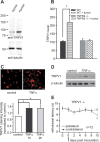
Heat hyperalgesia in the mice with tumor depended on TRPV1. A, Western blot analysis of TRPV1 expression in L3, L4, and L5 DRG neurons from control and tumor mice. TRPV1 protein level is 2.1-fold increased in tumor compared with naive mice. B, TRPV1 levels are significantly increased in tumor-bearing wild-type (n = 6) but not TNFR2−/− (n = 3) mice. C, Immunocytochemistry illustrates an increase in TRPV1 levels after TNFα (10 ng/ml) stimulation of DRG neurons in culture. Scale bar, 50 μm. The intensity of TRPV1 labeling increased significantly 1 and 2 h after TNFα (10 ng/ml) stimulation (p < 0.05 compared with control, ANOVA). D, TRPV1 protein level as detected by Western blot analysis was upregulated after TNFα (10 ng/ml) stimulation. E, Mice lacking the TRPV1 gene developed heat hyperalgesia only on day 10 after tumor induction (n = 12). Asterisks mark significant differences (p < 0.05, ANOVA). WT, Wild type.

References

    1. Alexander RB, Ponniah S, Hasday J, Hebel JR. Elevated levels of proinflammatory cytokines in the semen of patients with chronic prostatitis/chronic pelvic pain syndrome. Urology. 1998;52:744–749. - PubMed
    1. Baumgartner SW, Fleischmann RM, Moreland LW, Schiff MH, Markenson J, Whitmore JB. Etanercept (Enbrel) in patients with rheumatoid arthritis with recent onset versus established disease: improvement in disability. J Rheumatol. 2004;31:1532–1537. - PubMed
    1. Bhave G, Zhu W, Wang H, Brasier DJ, Oxford GS, Gereau RW. cAMP-dependent protein kinase regulates desensitization of the capsaicin receptor (VR1) by direct phosphorylation. Neuron. 2002;35:721–731. - PubMed
    1. Blumer MJ, Gahleitner P, Narzt T, Handl C, Ruthensteiner B. Ribbons of semithin sections: an advanced method with a new type of diamond knife. J Neurosci Methods. 2002;120:11–16. - PubMed
    1. Cain DM, Wacnik PW, Simone DA. Animal models of cancer pain may reveal novel approaches to palliative care. Pain. 2001a;91:1–4. - PubMed

Publication types

MeSH terms