

Review
doi: 10.1056/NEJMra0706596.
Tumor angiogenesis
Affiliations
- PMID: 18463380
- PMCID: PMC4542009
- DOI: 10.1056/NEJMra0706596
Item in Clipboard
Review
Tumor angiogenesis
N Engl J Med.
.
No abstract available
Figures
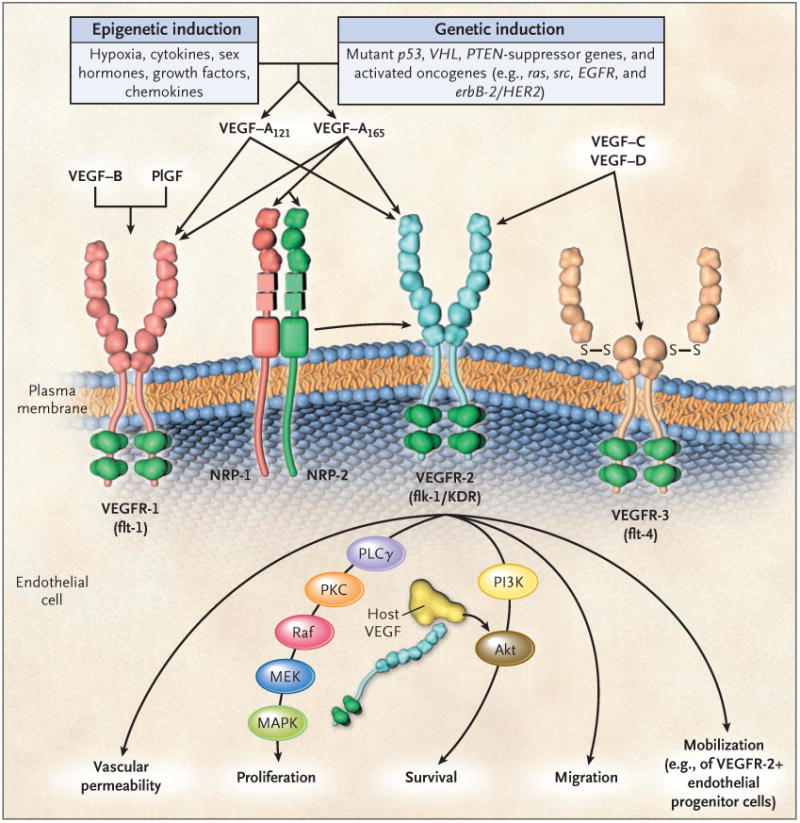
The major mediator of tumor angiogenesis is vascular endothelial growth factor A (VEGF-A, also called VEGF), specifically the circulating isoforms of VEGF — VEGF121 and VEGF165. These isoforms signal through VEGF receptor 2 (VEGFR-2), the major VEGF signaling receptor that mediates sprouting angiogenesis (called kinase-insert domain–containing receptor [KDR] in humans and fetal liver kinase 1 [flk-1] in mice). The role of VEGFR-1 in sprouting angiogenesis is much less clear. VEGF is expressed in most types of human cancer, and increased expression in tumors is often associated with a less favorable prognosis. Induction of or an increase in VEGF expression in tumors can be caused by numerous environmental (epigenetic) factors such as hypoxia, low pH, inflammatory cytokines (e.g., interleukin-6), growth factors (e.g., basic fibroblast growth factor), sex hormones (both androgens and estrogens), and chemokines (e.g., stromal-cell–derived factor 1). Other causes include genetic inductive changes such as activation of numerous different oncogenes or loss or mutational inactivation of a variety of tumor-suppressor genes. The binding of VEGF to VEGFR-2 leads to a cascade of different signaling pathways, two examples of which are shown, resulting in the up-regulation of genes involved in mediating the proliferation and migration of endothelial cells and promoting their survival and vascular permeability. For example, the binding of VEGF to VEGFR-2 leads to dimerization of the receptor, followed by intracellular activation of the PLCγ–PKC–Raf kinase–MEK–mitogen-activated protein kinase (MAPK) pathway and subsequent initiation of DNA synthesis and cell growth, whereas activation of the phosphatidylinositol 3′–kinase (PI3K)–Akt pathway leads to increased endothelial-cell survival. Activation of src can lead to actin cytoskeleton changes and induction of cell migration. VEGF receptors are located on the endothelial-cell surface; however, intracellular (“intracrine”)–signaling VEGF receptors (VEGFR-2) may be present as well, and they are involved in promoting the survival of endothelial cells. The detailed structure of the intracellular VEGFR-2 in endothelial cells is not yet known, but it is shown as the full-length receptor that is normally bound to the cell surface. Binding of VEGF-C to VEGFR-3 mediates lymphangiogenesis. VEGF165 can bind to neuropilin (NRP) receptors, which can act as coreceptors with VEGFR-2 (horizontal arrow) to regulate angiogenesis. EGFR denotes epidermal growth factor receptor, flt-1 fms-like tyrosine kinase 1, PlGF placental growth factor, PTEN phosphatase and tensin homologue, S–S disulfide bond, and VHL von Hippel–Lindau.
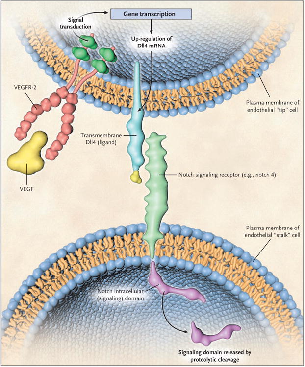
In mammals there are multiple deltalike ligands (Dlls), one of which, Dll4, is highly expressed in the vascular endothelial cells involved in angiogenesis. It tends to be expressed at higher levels in the “tip” cells of sprouting vessels and is induced by vascular endothelial growth factor (VEGF). Dll4 binds to notch receptors, two of which (notch 1 and notch 4) are expressed in the vascular endothelial cells composing the “stalk” component of a growing capillary sprout, adjacent to the tip cells. The interaction of Dll4 and notch receptors through the contact of adjacent endothelial cells leads to a series of proteolytic events whereby a notch intracellular signaling domain is cleaved and released by a γ-secretase; the domain then translocates to the nucleus. There it interacts with transcription factors and induces the expression of various target genes. The induction of Dll4–notch signaling is thought to act as a damping mechanism to prevent excessive angiogenesis and to promote the orderly development of new blood vessels. Blockade of Dll4–notch signaling interferes with this negative feedback mechanism, resulting in an increased density of vascular sprouts and branches, but these blood vessels are highly abnormal and do not perfuse blood adequately, leading to major increases in tumor hypoxia. The combined use of an anti-VEGF drug and a Dll4-targeting drug can be more effective than either drug used alone, and tumors that are resistant to an anti-VEGF drug can be treated with a Dll4-targeting drug. Thurston et al. describe more molecular details of Dll–notch receptor signaling and the domain structure of the receptor. VEGFR-2 denotes vascular endothelial growth factor receptor 2.
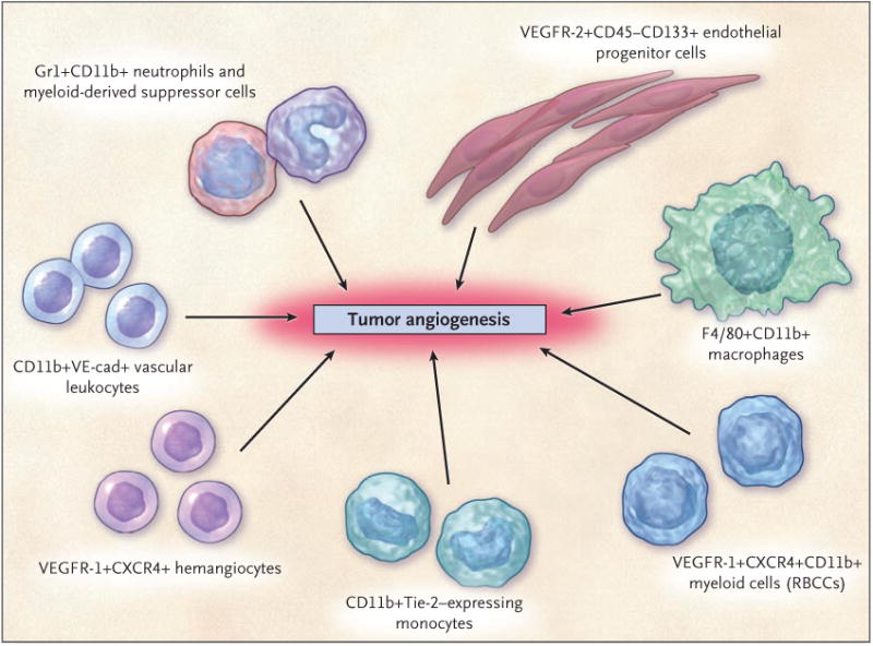
The various hematopoietic (CD45+) cell types appear to have a perivascular location with respect to the tumor neovasculature, whereas the CD45− endothelial progenitor cells can become incorporated into the lumen of a growing blood vessel and differentiate into mature endothelial cells. In recent preclinical studies, neutrophils have also been shown to contribute to the induction of tumor angiogenesis. F4/80 is a pan macrophage cell-surface marker. CXCR4 denotes CXC chemokine receptor 4, RBCCs recruited bone marrow–derived circulating cells, VE-cad vascular endothelial-cell cadherin (an adhesion molecule), and VEGFR vascular endothelial growth factor receptor. (Adapted from a figure provided by Dr. Michele dePalma, San Raffaele Scientific Institute, Milan.)
Comment in
-
Tumor angiogenesis.N Engl J Med. 2008 Aug 14;359(7):763; author reply 764. doi: 10.1056/NEJMc081278. N Engl J Med. 2008. PMID: 18703484 No abstract available.
References
-
- Folkman J. Tumor angiogenesis: therapeutic implications. N Engl J Med. 1971;285:1182–6. - PubMed
-
- Idem. Angiogenesis: an organizing principle for drug discovery? Nat Rev Drug Discov. 2007;6:273–86. - PubMed
-
- Ferrara N, Hillan KJ, Gerber HP, Novotny W. Discovery and development of bevacizumab, an anti-VEGF antibody for treating cancer. Nat Rev Drug Discov. 2004;3:391–400. - PubMed
-
- Hurwitz H, Fehrenbacher L, Novotny W, et al. Bevacizumab plus irinotecan, fluorouracil, and leucovorin for metastatic colorectal cancer. N Engl J Med. 2004;350:2335–42. - PubMed
-
- Sandler A, Gray R, Perry MC, et al. Paclitaxel–carboplatin alone or with bevacizumab for non–small-cell lung cancer. N Engl J Med. 2006;355:2542–50. Erratum, N Engl J Med 2007;356:318. - PubMed
Publication types
MeSH terms
Substances
Grants and funding
LinkOut - more resources
Full Text Sources
Other Literature Sources
Medical