. 2008 Jun 5;453(7196):745-50.
doi: 10.1038/nature07005. Epub 2008 May 7.

The branching programme of mouse lung development

Affiliations

The branching programme of mouse lung development

Ross J Metzger et al. Nature. .

Abstract

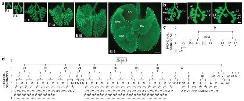
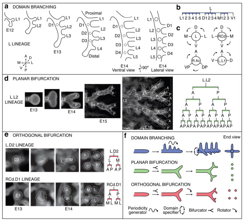
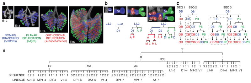
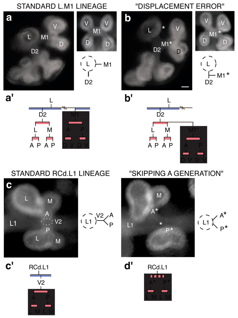
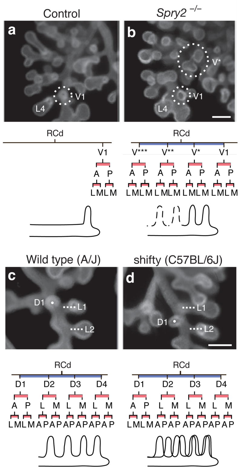
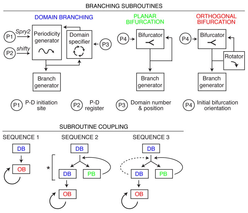
Mammalian lungs are branched networks containing thousands to millions of airways arrayed in intricate patterns that are crucial for respiration. How such trees are generated during development, and how the developmental patterning information is encoded, have long fascinated biologists and mathematicians. However, models have been limited by a lack of information on the normal sequence and pattern of branching events. Here we present the complete three-dimensional branching pattern and lineage of the mouse bronchial tree, reconstructed from an analysis of hundreds of developmental intermediates. The branching process is remarkably stereotyped and elegant: the tree is generated by three geometrically simple local modes of branching used in three different orders throughout the lung. We propose that each mode of branching is controlled by a genetically encoded subroutine, a series of local patterning and morphogenesis operations, which are themselves controlled by a more global master routine. We show that this hierarchical and modular programme is genetically tractable, and it is ideally suited to encoding and evolving the complex networks of the lung and other branched organs.


Figures

Figure 1
Figure 1. Branching morphogenesis of the mouse bronchial tree
a, Whole mount lungs (ventral view) at embryonic day indicated immunostained for E-Cadherin (green) to show airway epithelium. Dotted lines, right cranial (RCr), right middle (RMd), accessory (RAc), right caudal (RCd), and left (L) lobes. Bar, 500 μm. b, Reconstructing branching dynamics using three E12 specimens ~3 hours apart in age. Lateral 2° branches L1-3 (dots in b, c) sprout in proximal-to-distal order from left (L) 1° branch, as do lateral 2° branches L1 (box in b, c) and L2 from distal (RCd) portion of right (R) 1° branch. Bar, 200 μm. c, Branch lineage diagram for oldest lung in b. Branch names indicate lineage, e.g. RCd.L1 is first lateral 2° branch off RCd. d, Lineage diagram of RCd.L1 showing 250 descendant branches at E15 (box in a). A, anterior; D, dorsal; L, lateral; M, medial; P, posterior; V, ventral; *, orientation can vary.
Figure 2
Figure 2. Branching modes in lung development
a–c, Domain branching. a, Schematics of lateral and dorsal 2° branches budding from L. Lateral 2° branches (L1-5) bud in proximal to distal order. Proximal-to-distal branching begins again in second domain (projecting into plane of figure) to form a row of dorsal 2° branches (D1-4; dashed circles). Right panel, E14 schematic rotated 90° to show dorsal branches. b, Lineage diagram of 2° branches from L. Branches form in four domains: lateral (L), dorsal (D), medial (M), and ventral (V), indicated by blue bars. c, Schematic cross sections through L and three other branches indicated showing positions of domains and order (arrows) domains are used. d, Planar bifurcation. Ventral view of branch L.L2 in series of fixed specimens from E13 to E16 showing sequential bifurcations along A-P axis. E15 and E16 specimens were stained with anti-Smooth Muscle α-Actin to highlight early branch generations, which are surrounded by smooth muscle. Dotted lines outline bifurcations. Right panel, lineage of L.L2 descendants formed by planar bifurcation; branches not yet formed in E16 specimen are in gray. Bar, 100 μm. e, Orthogonal bifurcation. End-on (dorsal) views of branches indicated in developmental series of E13 and E14 specimens. L.D2 bifurcates along L-M axis, and its daughters along A-P axis, whereas RCd.D1 bifurcates along A-P axis and its daughters along L-M axis. Bar, 100 μm. f, Schematics of branching modes. The first bifurcation in a series is classified retrospectively, based on the orientation of the subsequent bifurcation. Icons show patterning and morphogenesis operations inferred for each mode: proximal-distal periodicity generator, circumferential domain specifier, branch bifurcator, and bifurcation plane rotator.
Figure 3
Figure 3. Deployment of branching modes
a, Lungs from Fig. 1a with branches pseudocolored blue (domain branching), green (planar bifurcation), or red (orthogonal bifurcation) to indicate branching mode used to form the branch. b, Close-up of L.L2 (dorsal view, lateral at right) pseudocolored as in a. Lineage diagrams are also colored to indicate branching mode. Branching mode can switch between generations and a single branch (e.g. L.L2) can form daughters by more than one mode. Bar, 100 μm. c, Sequences of branching mode use. DB, domain branching; OB, orthogonal bifurcation; PB, planar bifurcation. Sequence 2 includes a lineage formed by Sequence 1 (dotted box) and Sequence 3 includes a lineage formed by Sequence 2 (dotted box). d, Diagram showing sequence in c used to generate each lineage (named after the founder branch, see Supplementary Fig. 1) off the lobar branches (R.Cr, R.Md, R.Ac, RCd, L). Most or all lineages within a domain use the same sequence.
Figure 4
Figure 4. Branching errors
(a, b) “Displacement error”. a, Standard arrangement of D2 and M1 2° branches off L. Left panel, end-on (anterior) view of L and descendants. Right panel, end-on (medial) view of M1 and descendants; below, schematic of anterior view. a′, Standard lineage of L, D2 and M1. Grey box, M1 lineage. b, Same views as a of lung in which M1* arises off D2, normally a sister branch. Bar (for a and b), 50 μm. b, M1* follows the normal M1 lineage (grey box). (c, d) “Skipping a generation”. c, Standard arrangement of V2 arising off RCd.L1. Left panel, end-on (ventral) view of V2 and its descendants, which form by orthogonal bifurcation. Right panel, schematic depicting side (lateral) view of V2 and its daughters. c′, Standard lineage of RCd.L1 and V2. Grey box, descendants of V2. d, Same view (left) and schematic (right) of lung that skipped V2 generation. Bar (for c and d), 50 μm. Anterior (A*) and posterior (P*) branches sprout directly from RCd.L1, normally their grandparent. d′, Descendants of missing V2 (grey box) follow normal lineage.
Figure 5
Figure 5. Genetic control of branch pattern and lineage
(a, b) Ectopic domain branching in Spry2−/− mutants. a, RCd lobe (ventral view) of E12.5 control (Spry2+/−) lung with a single 2v branch (V1, circled) off RCd, at level of RCd.L4 (L4). Below, RCd.V1 lineage and schematic of RCd with single ventral 2° branch (V1). b, Same view of E12.5 Spry2−/− lung showing the normal ventral 2° branch (V1) and an ectopic branch (V*) that forms earlier and proximal to V1. V* has already sprouted additional generations of branches. Below, lineage and schematic show V* plus additional ectopic ventral branches (V**, V***; dashed lines in schematic) seen in other Spry2−/− lungs (Supplementary Fig. 4). Bar (for a and b), 200 μm. (c, d) Shifted domains in strain C57BL/6J. c, RCd lobe (dorsal view) of E12.5 lung from control strain A/J. Dorsal 2° branch RCd.D1 (D1; white dot) forms just proximal to lateral 2° branch RCd.L1 (L1, white line). Below, lineage and schematic of RCd.D1 and other branches in D domain. d, Same view of E12.5 lung from C57BL/6J mouse with shifty phenotype. RCd.D1 forms distal to RCd.L1. In shifty lungs, entire dorsal domain (black line in schematic) is shifted distally along RCd relative to wild type (grey line in schematic) but lineage is unaffected (except when there is a full unit shift and RCd.D4 lineage is missing, as indicated in gray; see Supplementary Fig. 5). Bar (for c and d), 200 μm.
Figure 6
Figure 6. A formal model of the lung branching program
(Top) Representation of branching modes as subroutines using different combinations of patterning and morphogenesis operations (boxed; see Fig. 2f). Subroutines are locally modified by input parameters P1–P4 that regulate variables indicated. P-D, proximal-distal. (Bottom) Three subroutine coupling schemes generate the three observed sequences of subroutine use (Fig. 3c). The schemes are related: bypass of step marked by asterisk in Sequence 2 generates Sequence 1, and repeat of domain branching step (dashed line) generates Sequence 3. DB, domain branching; PB, planar bifurcation; OB, orthogonal bifurcation.

Comment in

References

    1. Weibel ER. The pathway for oxygen. Harvard University Press, Cambridge; Massachusetts: 1984.
    1. West GB, Brown JH, Enquist BJ. A general model for the origin of allometric scaling laws in biology. Science. 1997;276:122–126. - PubMed
    1. Bejan A. Shape and structure, from engineering to nature. Cambridge University Press; Cambridge: 2000.
    1. Mauroy B, Filoche M, Weibel ER, Sapoval B. An optimal bronchial tree may be dangerous. Nature. 2004;427:633–636. - PubMed
    1. Aeby C. Der Bronchialbaum der Säugethiere und des Menschen, nebst Bemerkungen über den Bronchialbaum der Vögel und Reptilien. Engelmann; Leipzig: 1880.

Publication types

MeSH terms

Substances