. 2008 Oct;15(19):1311-20.
doi: 10.1038/gt.2008.78. Epub 2008 May 8.

Correction of the disease phenotype in the mouse model of Stargardt disease by lentiviral gene therapy

Affiliations

Correction of the disease phenotype in the mouse model of Stargardt disease by lentiviral gene therapy

J Kong et al. Gene Ther. 2008 Oct.

Abstract

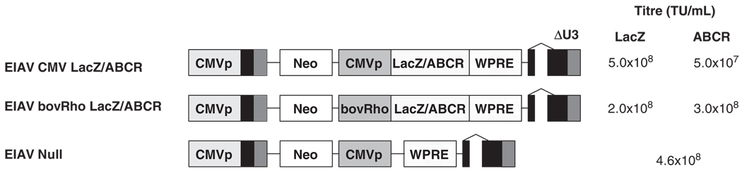
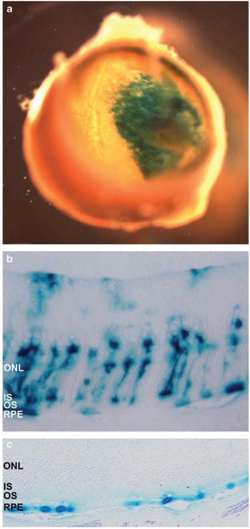
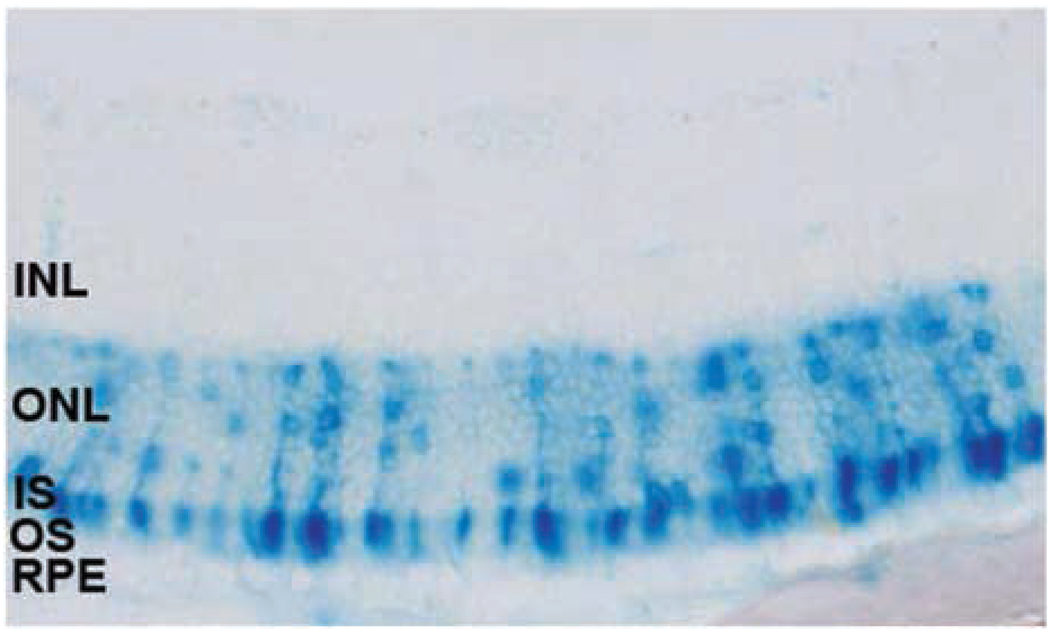
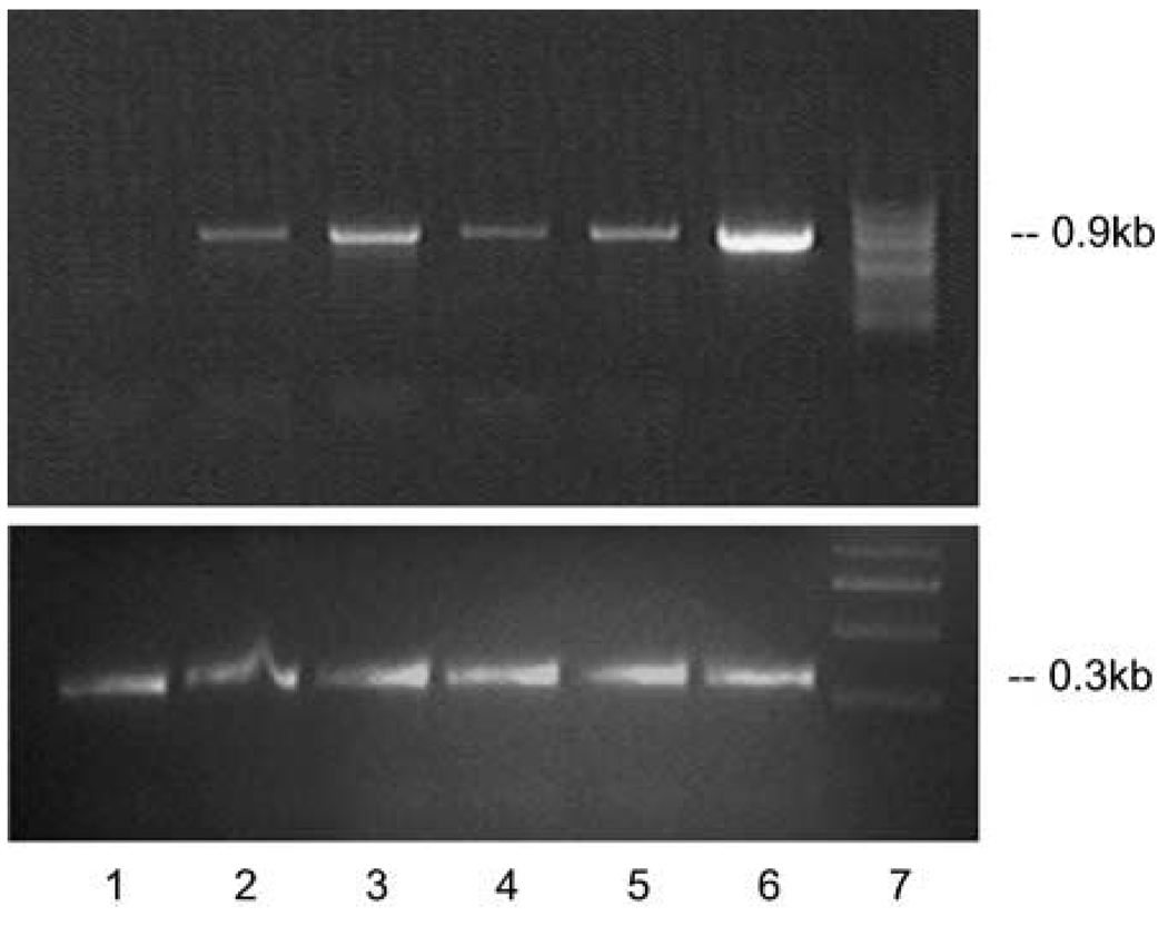
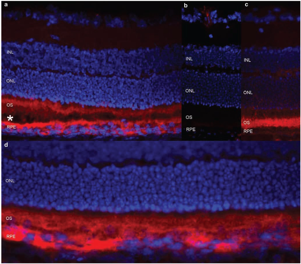
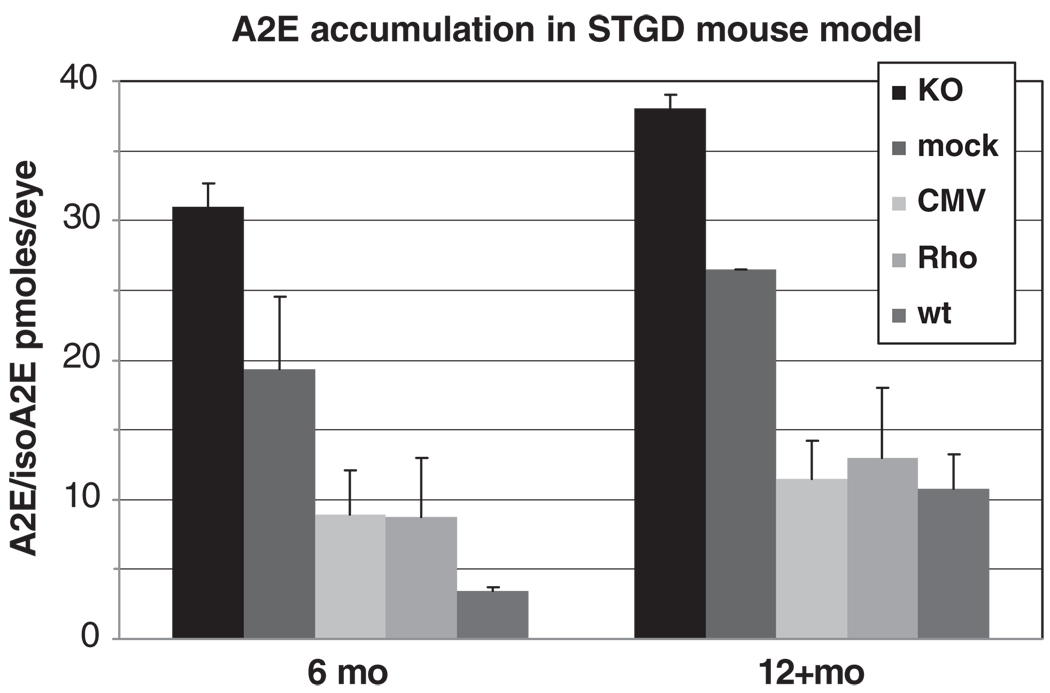
Autosomal recessive Stargardt disease (STGD1) is a macular dystrophy caused by mutations in the ABCA4 (ABCR) gene. The disease phenotype that is most recognized in STGD1 patients, and also in the Abca4-/- mouse (a disease model), is lipofuscin accumulation in retinal pigment epithelium. Here, we tested whether delivery of the normal (wt) human ABCA4 gene to the subretinal space of the Abca4 -/- mice via lentiviral vectors would correct the disease phenotype; that is, reduce accumulation of the lipofuscin pigment A2E. Equine infectious anemia virus (EIAV)-derived lentiviral vectors were constructed expressing either the human ABCA4 gene or the LacZ reporter gene under the control of the constitutive (CMV) or photoreceptor-specific (Rho) promoters. Abca4-/- mice were injected subretinally with 1 microl ( approximately 5.0 x 10(5) TU) of each EIAV vector in one eye at postnatal days 4 and 5. An injection of saline, an EIAV-null vector, or an uninjected contralateral eye served as a control. Mice were killed at various times after injection to determine photoreceptor (PR) transduction efficiency and A2E concentrations. EIAV-LacZ vectors transduced from 5 to 20% of the PRs in the injected area in mice. Most importantly, a single subretinal injection of EIAV-CMV-ABCA4 to Abca4-/- mouse eyes substantially reduced disease-associated A2E accumulation compared to untreated and mock-treated control eyes. Treated eyes of Abca4-/- mice accumulated 8-12 pmol per eye (s.d.=2.7) of A2E 1 year after treatment, amounts comparable to wt controls, whereas mock-treated or untreated eyes had 3-5 times more A2E (27-39 pmol per eye, s.d.=1.5; P=0.001-0.005). Although extrapolation to humans requires caution, the high transduction efficiency of both rod and cone photoreceptors and the statistically significant reduction of A2E accumulation in the mouse model of STGD1 suggest that lentiviral gene therapy is a potentially efficient tool for treating ABCA4-associated diseases.


Figures

Figure 1
Figure 1
Schematic representation of the equine infectious anemia virus (EIAV) vectors used in this study including the titer of each vector in transducing units per ml (TU ml−1). CMVp, the cytomegalovirus immediate/early promoter; Neo, Neomycin open reading frame; bovRho, 0.3 kb fragment from the bovine rhodopsin promoter; LacZ, β-galactosidase reporter gene, ABCR, human ABCR/ABCA4 cDNA; WPRE, modified Woodchuck hepatitis virus post-transcriptional regulatory element.
Figure 2
Figure 2
X-gal staining of the posterior segment (a) and Epon-embedded sections of mouse retinas transduced with the EIAV-CMV-LacZ vector (b) or the HIV-EF1α-LacZ vector (c). (a) A single injection of the EIAV-CMV-LacZ vector transduced ~20–30% of the entire retina. (b and c) Retinal sections of 18 days old mice, injected subretinally with 1 µl of viral solution containing 3–5 × 10⁵ TU of either EIAV-CMV-LacZ (b) or HIV-EF1α-LacZ (c) 14 days earlier. EIAV-derived vector transduced ~5% of photoreceptors, retinal pigment epithelium (RPE) and occasionally Mueller cells (b), whereas transduction was restricted to RPE cells with the HIV-derived vector (c). Representative images of n = 10 eyes (a and b) and n = 6 eyes (c) are shown.
Figure 3
Figure 3
X-gal staining of Epon section of a mouse retina following subretinal delivery of the equine infectious anemia virus (EIAV)-bRho-LacZ vector. The newborn mice (P4–5) were subretinally injected with 2.0 × 10⁵ TU of the EIAV-bRho-LacZ virus and X-gal staining was carried out 14 days later. The X-gal staining was restricted to the photoreceptor cell layer in all animals (n = 5).
Figure 4
Figure 4
Reverse transcriptase (RT)–PCR on the total RNA from eyecups of Abca4−/− mice treated with equine infectious anemia virus (EIAV)-ABCA4 vectors 2 weeks (lanes 2, 3) and 2 months (lanes 4–6) after subretinal injections. Upper panel: lane 1, untreated Abca4−/− mouse eyes; lane 2, Abca4−/− mouse eyes 2 weeks after EIAV-CMV-ABCR injection; lane 3, Abca4−/− mouse eyes 2 weeks after EIAV-bRho-ABCR injection; lane 4, Abca4−/− mouse eyes 2 months after EIAV-CMV-ABCR injection; lane 5, the same experiment as in lane 4 with a different isolate of the EIAV-CMV-ABCR virus; lane 6, Abca4−/− mouse eyes 2 months after EIAV-bRho-ABCR injection; lane 7, marker. Lower panel shows amplification of a 0.3 kb fragment of the actin gene from the same RNA isolates.
Figure 5
Figure 5
Expression of the human and murine ABCA4 protein in the mouse retina. (a) The Abca4−/− mouse retina treated with the EIAV-CMV-ABCA4 virus, showing the human ABCA4 in photoreceptor (PR) outer segments (OS) and in the RPE (in red). OS and RPE cell layers are slightly detached (shown by *) to further separate the staining in the PR outer segments and RPE cells. (b) Untreated Abca4−/− mouse, lacking any ABCA4/Abca4. (c) Wild-type mouse showing the endogenous mouse Abca4 protein in PR outer segments. (d) The same experiment as in (a), showing the extent of the expression of the human ABCA4 protein in the treated area with no detachment. INL, inner nuclear layer; ONL, outer nuclear layer; OS, outer segments of photoreceptors; RPE, retinal pigment epithelium.
Figure 6
Figure 6
A2E/isoA2E content in mouse eyes at 6 and 12+ months time points. KO, untreated Abca4−/− mice; mock, Abca4−/− animals injected with equine infectious anemia virus (EIAV)-null virus; CMV, Abca4−/− mice treated with EIAV-CMV-ABCA4 construct; Rho, Abca4−/− mice treated with EIAV-Rho-ABCA4 constructs; wt, wild type animals. Each bar (data point) represents n = 4–20 eyes with s.d. values shown.

References

    1. Blacharski P. In: Retinal dystrophies and degenerations. Newsome DA, editor. New York: Raven Press; 1988. pp. 135–159.
    1. Anderson KL, Baird L, Lewis RA, Chinault AC, Otterud B, Leppert M, et al. A YAC contig encompassing the recessive Stargardt disease gene (STGD) on chromosome 1p. Am J Hum Genet. 1995;57:1351–1363. - PMC - PubMed
    1. Stargardt K. Über familiäre, progressive Degeneration in der Maculagegend des Auges. Albrecht von Graefes Arch Ophthalmol. 1909;71:534–550.
    1. Allikmets R, Singh N, Sun H, Shroyer NF, Hutchinson A, Chidambaram A, et al. A photoreceptor cell-specific ATP-binding transporter gene (ABCR) is mutated in recessive Stargardt macular dystrophy. Nat Genet. 1997a;15:236–246. - PubMed
    1. Azarian SM, Travis GH. The photoreceptor rim protein is an ABC transporter encoded by the gene for recessive Stargardt’s disease (ABCR) FEBS Lett. 1997;409:247–252. - PubMed

Publication types

MeSH terms

Substances