

Percutaneous CT-guided biopsy of the spine: results of 430 biopsies
- PMID: 18463900
- PMCID: PMC2443265
- DOI: 10.1007/s00586-008-0678-x
Percutaneous CT-guided biopsy of the spine: results of 430 biopsies
Abstract
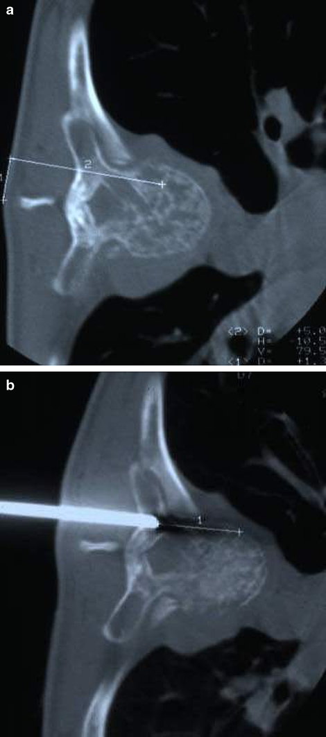
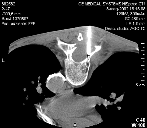
Biopsies of lesions in the spine are often challenging procedures with significant risk of complications. CT-guided needle biopsies could lower these risks but uncertainties still exist about the diagnostic accuracy. Aim of this retrospective study was to evaluate the diagnostic accuracy of CT-guided needle biopsies for bone lesions of the spine. We retrieved the results of 430 core needle biopsies carried out over the past fifteen years at the authors' institute and examined the results obtained. Of the 430 biopsies performed, in 401 cases the right diagnosis was made with the first CT-guided needle biopsy (93.3% accuracy rate). Highest accuracy rates were obtained in primary and secondary malignant lesions. Most false negative results were found in cervical lesions and in benign, pseudotumoral, inflammatory, and systemic pathologies. There were only 9 complications (5 transient paresis, 4 haematomas that resolved spontaneously) that had no influence on the treatment strategy, nor on the patient's outcome. In conclusion we can assert that this technique is reliable and safe and should be considered the gold standard in biopsies of the spine.
Figures

References
-
- Ayala AG, Ro JY, Fanning CV, Flores JP, Yasko AW. Core needle biopsy and fine needle aspiration in the diagnosis of bone and soft tissue lesions. Hematol Oncol Clin North Am. 1995;9:633–651. - PubMed
-
- Bickels J, Jelinek J, Shmookler B, Neff RS, Malawer MM. Biopsy of muscoluloskeletal tumors. Current concepts. Cinical Orthop Relat Res. 2001;368:212–219. - PubMed
-
- Campanacci M, Mercuri M, Gamberini G. Biopsy. Chir Organi Mov. 1995;80(2):113–123. - PubMed
-
- Dondelinger RF, Vanderschelden P, Capasso P, Trotteur G. Spiral tomodensitometry applied to interventional procedures. J Belge Radiol. 1995;78:118–125. - PubMed
MeSH terms
LinkOut - more resources
Full Text Sources
Medical
Miscellaneous
