. 2008 May 8;453(7192):184-9.
doi: 10.1038/nature06941.

Dynamic binding orientations direct activity of HIV reverse transcriptase

Affiliations

Dynamic binding orientations direct activity of HIV reverse transcriptase

Elio A Abbondanzieri et al. Nature. .

Abstract

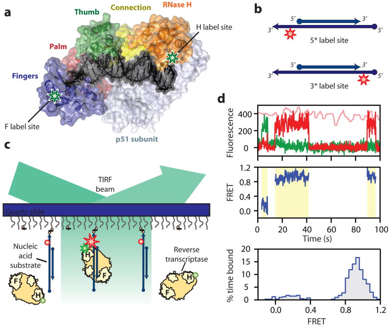
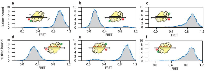
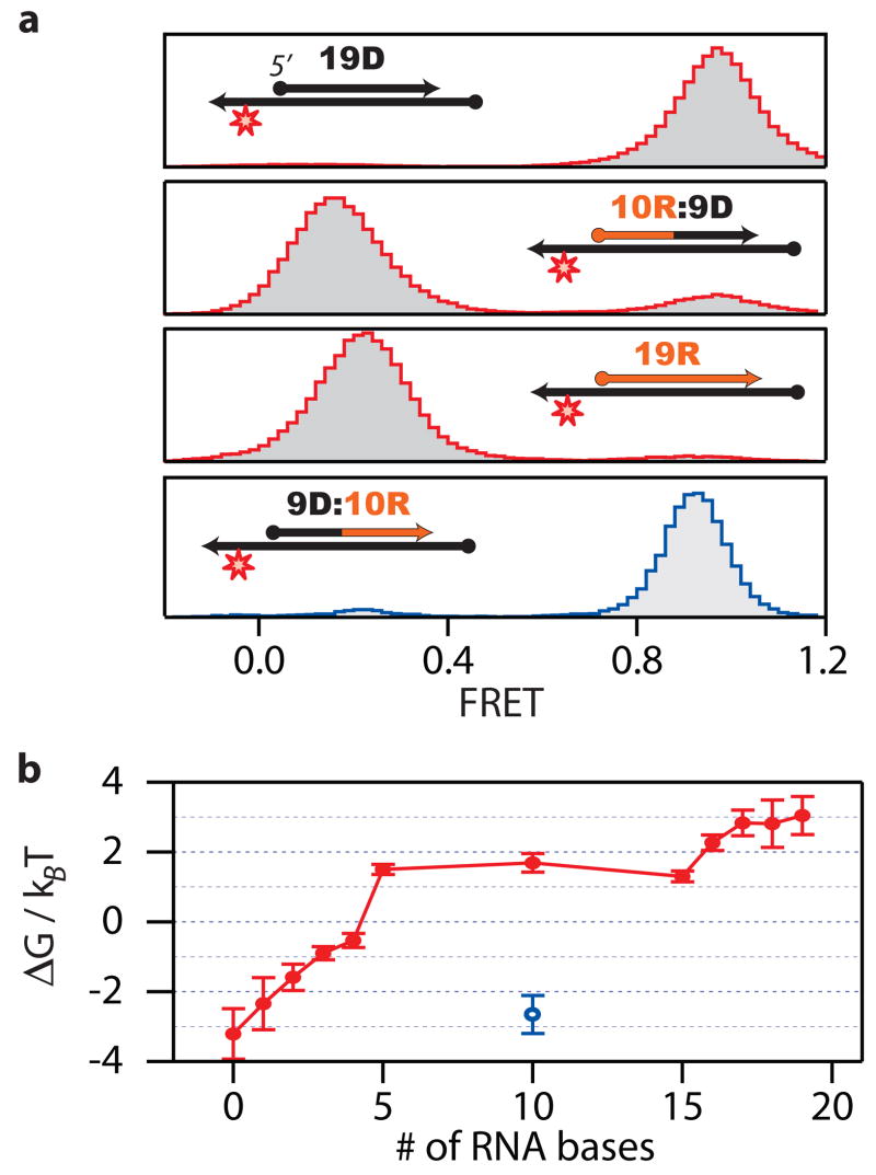
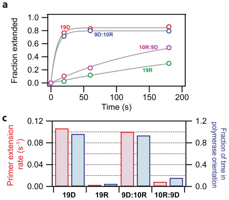
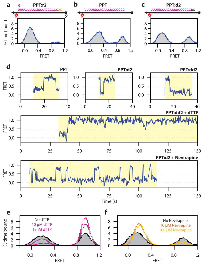
The reverse transcriptase of human immunodeficiency virus (HIV) catalyses a series of reactions to convert the single-stranded RNA genome of HIV into double-stranded DNA for host-cell integration. This task requires the reverse transcriptase to discriminate a variety of nucleic-acid substrates such that active sites of the enzyme are correctly positioned to support one of three catalytic functions: RNA-directed DNA synthesis, DNA-directed DNA synthesis and DNA-directed RNA hydrolysis. However, the mechanism by which substrates regulate reverse transcriptase activities remains unclear. Here we report distinct orientational dynamics of reverse transcriptase observed on different substrates with a single-molecule assay. The enzyme adopted opposite binding orientations on duplexes containing DNA or RNA primers, directing its DNA synthesis or RNA hydrolysis activity, respectively. On duplexes containing the unique polypurine RNA primers for plus-strand DNA synthesis, the enzyme can rapidly switch between the two orientations. The switching kinetics were regulated by cognate nucleotides and non-nucleoside reverse transcriptase inhibitors, a major class of anti-HIV drugs. These results indicate that the activities of reverse transcriptase are determined by its binding orientation on substrates.


Figures

Figure 1
Figure 1. Single-molecule FRET assay for probing the orientational dynamics of RT
a, The structure of HIV-1 RT bound to a DNA-DNA substrate. Labelling sites for Cy3 on RT are highlighted by green stars. b, Nucleic-acid substrates consisted of a 19–21 nt primer strand annealed to a 50 nt template strand containing an Cy5 label (red star). Cy5 was either 3 nt from the 5′ end (circle) or 4–6 nt from the 3′ end (arrow) of the primer. c, Single-molecule detection of Cy3 (green star or sphere) labelled RT binding to and dissociating from the surface-immobilized nucleic-acid substrates labelled with Cy5 (red star or sphere). The stars and spheres indicated dyes that do or do not emit fluorescence, respectively. d, FRET analysis for RT binding to a single primer-template complex: The upper panel shows the fluorescence time traces from Cy3 (green) and Cy5 (red) under 532 nm excitation and that from Cy5 (pink) under 635 nm excitation. The FRET value is calculated over the duration of the binding events (middle panel, yellow shaded regions) and a FRET distribution histogram is created for the binding events (lower panel).
Figure 2
Figure 2. FRET distributions of RT bound to nucleic-acid substrates reveal distinct RT binding orientations on RNA and DNA primers
a, RT with Cy3 (green star) attached in the H-labelling scheme was allowed to bind substrates consisting of DNA primer and template (black arrow), with Cy5 (red star) attached in the 5*-labelling scheme. b, H-labelled RT bound to 3*-labelled DNA duplex substrates. c, F-labelled RT bound to 3*-labelled DNA duplex substrates. d, H-labelled RT bound to 5*-labelled hybrid duplex substrates consisting of a RNA primer (orange arrow) and a DNA template (black arrow). e, H-labelled RT bound to 3*-labelled hybrid duplex substrates. f, F-labelled RT bound to 5*-labelled hybrid duplex substrates. The RT binding orientations consistent with the FRET distributions are depicted.
Figure 3
Figure 3. Binding orientation of RT on chimeric substrates
a, Selected FRET distributions of H-labelled RT bound to 5*-labelled substrates containing various 19 nt chimeric RNA:DNA primers hybridized to a DNA template. FRET distributions of other xR:yD substrates are shown in supplementary Fig. 7. b, The free energy difference ΔG between the high and low FRET orientations is plotted as a function of RNA content for both xR:yD (red) and 9D:10R (blue) chimeras. The error bars are the standard error of the mean (N = 3).
Figure 4
Figure 4. The DNA polymerase activity of RT correlates with its binding orientation on substrates
a, Primer extension activity of RT assayed on four selected primers (19D, 19R, 9D:10R and 10R:9D) annealed to a DNA template. The fraction of primers which had been extended by more than one bases is plotted as a function of time for the four primers (coloured circles). The data were fit to a single-exponential decay (grey lines) to deduce the primer extension rate constants. b, Rate constants of primer extension (red) correlate with the fractional of time the RT bound in the high FRET orientation conducive to polymerization (blue).
Figure 5
Figure 5. Dynamic binding orientations of RT on PPT substrates
a-c, FRET histograms of H-labelled RT bound to substrates containing 5*-labelled PPT:r2, PPT or PPT:d2 primers annealed to DNA templates. The PPT sequence is highlighted in violet letters and the 2 nt RNA and DNA extensions are coloured in orange and black, respectively. The DNA template is shown as a black arrow. d, FRET time traces of RT bound to PPT, PPT:d2, and PPT:dd2 substrates showed spontaneous flipping transitions between the two binding orientations. e, FRET histograms of RT bound to PPT:dd2 substrates in the presence of 0, 10 μM, and 1 mM dTTP. f, FRET histograms of RT bound to PPT:d2 substrates in the presence of 0, 10, and 100 μM Nevirapine.

Comment in

References

    1. Goff Stephen P. In: Fields Virology. Knipe DM, Howley PM, editors. Vol. 2. Lippincott Williams and Wilkins; Philadelphia: 2001. pp. 1871–1940.
    1. Champoux JJ. In: Reverse Transcriptase. Skalka AM, Goff SP, editors. Cold Spring Harbor Laboratory Press; New York: 1993. pp. 10–118.
    1. Hostomsky Z, Hostomska Z, Fu TB, Taylor J. Reverse transcriptase of human immunodeficiency virus type 1: functionality of subunits of the heterodimer in DNA synthesis. J Virol. 1992;66:3179–3182. - PMC - PubMed
    1. Kohlstaedt LA, Wang J, Friedman JM, Rice PA, Steitz TA. Crystal structure at 3.5 A resolution of HIV-1 reverse transcriptase complexed with an inhibitor. Science. 1992;256:1783–1790. - PubMed
    1. Baltimore D. Viral RNA-dependent DNA Polymerase: RNA-dependent DNA Polymerase in Virions of RNA Tumour Viruses. Nature. 1970;226:1209–1211. - PubMed

Publication types