

Fresh osteochondral allografts for posttraumatic knee defects: long-term followup
- PMID: 18465182
- PMCID: PMC2584250
- DOI: 10.1007/s11999-008-0282-8
Fresh osteochondral allografts for posttraumatic knee defects: long-term followup
Abstract
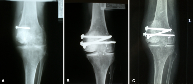
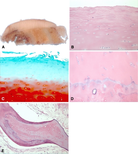
Fresh osteochondral allograft transplantation has been an effective treatment option with promising long-term clinical outcomes for focal posttraumatic defects in the knee for young, active individuals. We examined histologic features of 35 fresh osteochondral allograft specimens retrieved at the time of subsequent graft revision, osteotomy, or TKA. Graft survival time ranged from 1 to 25 years based on their time to reoperation. Histologic features of early graft failures were lack of chondrocyte viability and loss of matrix cationic staining. Histologic features of late graft failures were fracture through the graft, active and incomplete remodeling of the graft bone by the host bone, and resorption of the graft tissue by synovial inflammatory activity at graft edges. Histologic features associated with long-term allograft survival included viable chondrocytes, functional preservation of matrix, and complete replacement of the graft bone with the host bone. Given chondrocyte viability, long-term allograft survival depends on graft stability by rigid fixation of host bone to graft bone. With the stable osseous graft base, the hyaline cartilage portion of the allograft can survive and function for 25 years or more.
Figures
References
-
- {'text': '', 'ref_index': 1, 'ids': [{'type': 'DOI', 'value': '10.1097/00003086-200110001-00029', 'is_inner': False, 'url': 'https://doi.org/10.1097/00003086-200110001-00029'}, {'type': 'PubMed', 'value': '11603715', 'is_inner': True, 'url': 'https://pubmed.ncbi.nlm.nih.gov/11603715/'}]}
- Aubin PP, Cheah HK, Davis AM, Gross AE. Long-term follow-up of fresh osteochondral allografts for posttraumatic knee defects. Clin Orthop Relat Res. 2001;391:318–327. - PubMed
-
- {'text': '', 'ref_index': 1, 'ids': [{'type': 'DOI', 'value': '10.1007/s002640050260', 'is_inner': False, 'url': 'https://doi.org/10.1007/s002640050260'}, {'type': 'PMC', 'value': 'PMC3619576', 'is_inner': False, 'url': 'https://pmc.ncbi.nlm.nih.gov/articles/PMC3619576/'}, {'type': 'PubMed', 'value': '9914928', 'is_inner': True, 'url': 'https://pubmed.ncbi.nlm.nih.gov/9914928/'}]}
- Bakay A, Csonge L, Papp G, Fekete L. Osteochondral resurfacing of the knee joint with allograft: clinical analysis of 33 cases. Int Orthop. 1998;22:277–281. - PMC - PubMed
-
- {'text': '', 'ref_index': 1, 'ids': [{'type': 'DOI', 'value': '10.1302/0301-620X.87B3.15552', 'is_inner': False, 'url': 'https://doi.org/10.1302/0301-620x.87b3.15552'}, {'type': 'PubMed', 'value': '15773640', 'is_inner': True, 'url': 'https://pubmed.ncbi.nlm.nih.gov/15773640/'}]}
- Bartlett W, Gooding CR, Carrington RW, Skinner JA, Briggs TW, Bentley G. Autologous chondrocyte implantation at the knee using a bilayer collagen membrane with bone graft.: a preliminary report. J Bone Joint Surg Br. 2005;87:330–332. - PubMed
-
- {'text': '', 'ref_index': 1, 'ids': [{'type': 'PubMed', 'value': '3903605', 'is_inner': True, 'url': 'https://pubmed.ncbi.nlm.nih.gov/3903605/'}]}
- Bayne O, Langer F, Pritzker KP, Houpt J, Gross AE. Osteochondral allografts in the treatment of osteonecrosis of the knee. Orthop Clin North Am. 1985;16:727–740. - PubMed
-
- {'text': '', 'ref_index': 1, 'ids': [{'type': 'PubMed', 'value': '1732235', 'is_inner': True, 'url': 'https://pubmed.ncbi.nlm.nih.gov/1732235/'}]}
- Beaver RJ, Mahomed M, Backstein D, Davis A, Zukor DJ, Gross AE. Fresh osteochondral allografts for post-traumatic defects in the knee: a survivorship analysis. J Bone Joint Surg Br. 1992;74:105–110. - PubMed
MeSH terms
LinkOut - more resources
Full Text Sources
Other Literature Sources
Medical
