

White matter hyperintensities in the forties: their prevalence and topography in an epidemiological sample aged 44-48
- PMID: 18465744
- PMCID: PMC6870596
- DOI: 10.1002/hbm.20586
White matter hyperintensities in the forties: their prevalence and topography in an epidemiological sample aged 44-48
Abstract
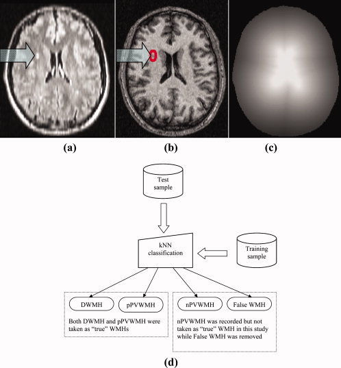
White matter hyperintensities (WMHs) are a frequent finding on T2-weighted MRI of the brain in elderly individuals, but their prevalence and severity in younger asymptomatic populations is less well studied. We report the topography of WMHs on T2-weighted fluid inversion recovery (FLAIR) MRI in 428 individuals aged 44-48 years recruited randomly from a healthy community sample. WMHs were delineated from FLAIR and T1-weighted scans by using a computer algorithm, further verified and then classified using k-nearest neighbor (kNN) algorithm into deep WMH (DWMH), and periventricular WMH (PVWMH), which included extended periventricular "rims" and frontal and occipital "caps". Small caps and pencil-thin rims were not taken as WMHs for this analysis. The new computer algorithm was validated and compared with the scores of visual rating, and the correspondence between the two methods was high. We found that 218 (50.9%) subjects had WMHs. 146 of the 218 (34.1% of whole sample population of 428) subjects had deep white matter hyperintensities (DWMHs). The average number of WMH clusters (occurrences) per brain was 1.37 (0.94 for DWMH and 0.43 for pathological PVWMH) and the mean WMH tissue volume was 0.278 ml. There was no significant sex difference in the severity and distribution of WMHs. The study suggests that small punctate or focal WMHs are common in the brains of individuals in their 40s, and may represent an early stage of development of these lesions.
2008 Wiley-Liss, Inc.
Figures
References
-
- Altaf N,Daniels L,Morgan PS,Lowe J,Gladman J,MacSweeney ST,Moody A,Auer DP ( 2006): Cerebral white matter hyperintense lesions are associated with unstable carotid plaques. Eur J Vasc Endovasc Surg 31: 8–13. - PubMed
-
- Anbeek P,Vincken KL,van Osch MJP,Bisschops RHC,van der Grond J ( 2004): Automatic segmentation of different‐sized white matter lesions by voxel probability estimation. Med Image Anal 8: 205–215. - PubMed
-
- Ashburner J,Friston K ( 1997): Multimodal image coregistration and partitioning‐‐A unified framework. NeuroImage 6: 209–217. - PubMed
-
- Ashburner J,Friston KJ ( 2005): Unified segmentation. NeuroImage 26: 839–851. - PubMed
Publication types
MeSH terms
LinkOut - more resources
Full Text Sources
Medical
Miscellaneous
